 Movie
Movie Controller

[English] 日本語
 Yorodumi
- PDB-5uc9: Crystal structure of human Heme Oxygenase-2 in complex with Myristate -
+ Open data

- Basic information
| Entry | Database: PDB / ID: 5uc9 | ||||||
|---|---|---|---|---|---|---|---|
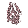
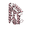
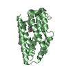
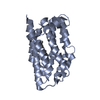
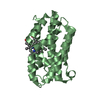
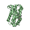
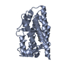
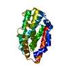
| Title | Crystal structure of human Heme Oxygenase-2 in complex with Myristate | ||||||
 Components Components | Heme oxygenase 2 | ||||||
 Keywords Keywords | OXIDOREDUCTASE / heme oxygenase / myristic acid | ||||||
| Function / homology |  Function and homology information Function and homology informationheme oxygenase (biliverdin-producing) / heme oxidation / heme oxygenase (decyclizing) activity / heme catabolic process / Heme degradation / RHOA GTPase cycle / specific granule membrane / Cytoprotection by HMOX1 / Iron uptake and transport / response to oxidative stress ...heme oxygenase (biliverdin-producing) / heme oxidation / heme oxygenase (decyclizing) activity / heme catabolic process / Heme degradation / RHOA GTPase cycle / specific granule membrane / Cytoprotection by HMOX1 / Iron uptake and transport / response to oxidative stress / response to hypoxia / heme binding / Neutrophil degranulation / endoplasmic reticulum membrane / membrane / metal ion binding / plasma membrane Similarity search - Function | ||||||
| Biological species |  Homo sapiens (human) Homo sapiens (human) | ||||||
| Method |  X-RAY DIFFRACTION / SYNCHROTRON / MOLECULAR REPLACEMENT / Resolution: 1.903 Å X-RAY DIFFRACTION / SYNCHROTRON / MOLECULAR REPLACEMENT / Resolution: 1.903 Å | ||||||
 Authors Authors | Luo, S. / Tong, L. | ||||||
 Citation Citation | Journal: Cell Host Microbe / Year: 2017 Title: Heme Oxygenase 2 Binds Myristate to Regulate Retrovirus Assembly and TLR4 Signaling. Authors: Zhu, Y. / Luo, S. / Sabo, Y. / Wang, C. / Tong, L. / Goff, S.P. | ||||||
| History |
|
- Structure visualization
| Structure viewer | Molecule: MolmilJmol/JSmol |
|---|
- Downloads & links
-Download
| PDBx/mmCIF format | 5uc9.cif.gz | 195.4 KB | Display | PDBx/mmCIF format |
|---|---|---|---|---|
| PDB format | pdb5uc9.ent.gz | 154.8 KB | Display | PDB format |
| PDBx/mmJSON format | 5uc9.json.gz | Tree view | PDBx/mmJSON format | |
| Others |  Other downloads Other downloads |
-Validation report
| Arichive directory | https://data.pdbj.org/pub/pdb/validation_reports/uc/5uc9ftp://data.pdbj.org/pub/pdb/validation_reports/uc/5uc9 | HTTPS FTP |
|---|
-Related structure data
| Related structure data | 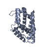 5uc8SC  5ucaC S: Starting model for refinement C: citing same article ( |
|---|---|
| Similar structure data |


-Links
 PDBj
PDBj


- Assembly
| Deposited unit | 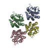
| ||||||||
|---|---|---|---|---|---|---|---|---|---|
| 1 | 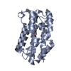
| ||||||||
| 2 | 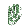
| ||||||||
| 3 | 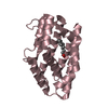
| ||||||||
| 4 | 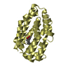
| ||||||||
| Unit cell |
|
-Components
| #1: Protein | Mass: 26286.639 Da / Num. of mol.: 4 / Fragment: residues 1-213 Source method: isolated from a genetically manipulated source Source: (gene. exp.) Homo sapiens (human) / Gene: HMOX2, HO2 / Production host:  References: UniProt: P30519, heme oxygenase (biliverdin-producing) #2: Chemical | ChemComp-MYR /   Mass: 228.371 Da / Num. of mol.: 4 / Source method: obtained synthetically / Formula: C14H28O2 Mass: 228.371 Da / Num. of mol.: 4 / Source method: obtained synthetically / Formula: C14H28O2#3: Water | ChemComp-HOH / |  Mass: 18.015 Da / Num. of mol.: 584 / Source method: isolated from a natural source / Formula: H2O Mass: 18.015 Da / Num. of mol.: 584 / Source method: isolated from a natural source / Formula: H2O |
|---|
-Experimental details
-Experiment
| Experiment | Method: X-RAY DIFFRACTION / Number of used crystals: 1 |
|---|
- Sample preparation
| Crystal | Density Matthews: 2.1 Å3/Da / Density % sol: 41.44 % |
|---|---|
| Crystal grow | Temperature: 293 K / Method: vapor diffusion Details: 0.1 M Bis-Tris (pH 6.5) and 24% (w/v) PEG 2,000 monomethyl ether |
-Data collection
| Diffraction | Mean temperature: 100 K | ||||||||||||||||||||||||||||||||||||||||||||||||||||||||||||||||||
|---|---|---|---|---|---|---|---|---|---|---|---|---|---|---|---|---|---|---|---|---|---|---|---|---|---|---|---|---|---|---|---|---|---|---|---|---|---|---|---|---|---|---|---|---|---|---|---|---|---|---|---|---|---|---|---|---|---|---|---|---|---|---|---|---|---|---|---|
| Diffraction source | Source: SYNCHROTRON / Site: APS  / Beamline: 24-ID-E / Wavelength: 0.97921 Å / Beamline: 24-ID-E / Wavelength: 0.97921 Å | ||||||||||||||||||||||||||||||||||||||||||||||||||||||||||||||||||
| Detector | Type: ADSC QUANTUM 315 / Detector: CCD / Date: Apr 10, 2015 / Details: LR-Design detector positioner | ||||||||||||||||||||||||||||||||||||||||||||||||||||||||||||||||||
| Radiation | Protocol: SINGLE WAVELENGTH / Monochromatic (M) / Laue (L): M / Scattering type: x-ray | ||||||||||||||||||||||||||||||||||||||||||||||||||||||||||||||||||
| Radiation wavelength | Wavelength: 0.97921 Å / Relative weight: 1 | ||||||||||||||||||||||||||||||||||||||||||||||||||||||||||||||||||
| Reflection | Resolution: 1.9→25 Å / Num. obs: 68092 / % possible obs: 97.3 % / Redundancy: 3.6 % / Biso Wilson estimate: 29.83 Å2 / Rmerge(I) obs: 0.058 / Rpim(I) all: 0.034 / Rrim(I) all: 0.067 / Χ2: 1.039 / Net I/σ(I): 14.1 / Num. measured all: 246452 | ||||||||||||||||||||||||||||||||||||||||||||||||||||||||||||||||||
| Reflection shell |
|
- Processing
| Software |
| |||||||||||||||||||||||||||||||||||||||||||||||||||||||||||||||||||||||||||||||||||||||||||||||||||||||||||||||||||||||||||||||||||||||||||||||||||||||||||||||||||||||||||||||||||||||||||||||||||||||||||||||||||||||||
|---|---|---|---|---|---|---|---|---|---|---|---|---|---|---|---|---|---|---|---|---|---|---|---|---|---|---|---|---|---|---|---|---|---|---|---|---|---|---|---|---|---|---|---|---|---|---|---|---|---|---|---|---|---|---|---|---|---|---|---|---|---|---|---|---|---|---|---|---|---|---|---|---|---|---|---|---|---|---|---|---|---|---|---|---|---|---|---|---|---|---|---|---|---|---|---|---|---|---|---|---|---|---|---|---|---|---|---|---|---|---|---|---|---|---|---|---|---|---|---|---|---|---|---|---|---|---|---|---|---|---|---|---|---|---|---|---|---|---|---|---|---|---|---|---|---|---|---|---|---|---|---|---|---|---|---|---|---|---|---|---|---|---|---|---|---|---|---|---|---|---|---|---|---|---|---|---|---|---|---|---|---|---|---|---|---|---|---|---|---|---|---|---|---|---|---|---|---|---|---|---|---|---|---|---|---|---|---|---|---|---|---|---|---|---|---|---|---|---|
| Refinement | Method to determine structure: MOLECULAR REPLACEMENT Starting model: 5UC8 Resolution: 1.903→25 Å / SU ML: 0.21 / Cross valid method: FREE R-VALUE / σ(F): 1.34 / Phase error: 25.08
| |||||||||||||||||||||||||||||||||||||||||||||||||||||||||||||||||||||||||||||||||||||||||||||||||||||||||||||||||||||||||||||||||||||||||||||||||||||||||||||||||||||||||||||||||||||||||||||||||||||||||||||||||||||||||
| Solvent computation | Shrinkage radii: 0.9 Å / VDW probe radii: 1.11 Å | |||||||||||||||||||||||||||||||||||||||||||||||||||||||||||||||||||||||||||||||||||||||||||||||||||||||||||||||||||||||||||||||||||||||||||||||||||||||||||||||||||||||||||||||||||||||||||||||||||||||||||||||||||||||||
| Displacement parameters | Biso max: 77.45 Å2 / Biso mean: 33.3945 Å2 / Biso min: 15.63 Å2 | |||||||||||||||||||||||||||||||||||||||||||||||||||||||||||||||||||||||||||||||||||||||||||||||||||||||||||||||||||||||||||||||||||||||||||||||||||||||||||||||||||||||||||||||||||||||||||||||||||||||||||||||||||||||||
| Refinement step | Cycle: final / Resolution: 1.903→25 Å
| |||||||||||||||||||||||||||||||||||||||||||||||||||||||||||||||||||||||||||||||||||||||||||||||||||||||||||||||||||||||||||||||||||||||||||||||||||||||||||||||||||||||||||||||||||||||||||||||||||||||||||||||||||||||||
| Refine LS restraints |
| |||||||||||||||||||||||||||||||||||||||||||||||||||||||||||||||||||||||||||||||||||||||||||||||||||||||||||||||||||||||||||||||||||||||||||||||||||||||||||||||||||||||||||||||||||||||||||||||||||||||||||||||||||||||||
| LS refinement shell | Refine-ID: X-RAY DIFFRACTION / Rfactor Rfree error: 0 / Total num. of bins used: 30
|
