 Movie
Movie Controller

[English] 日本語
 Yorodumi
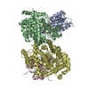
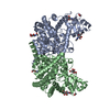
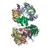
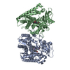
- PDB-1i9c: GLUTAMATE MUTASE FROM CLOSTRIDIUM COCHLEARIUM: COMPLEX WITH ADENO... -
+ Open data

- Basic information
| Entry | Database: PDB / ID: 1i9c | ||||||
|---|---|---|---|---|---|---|---|
| Title | GLUTAMATE MUTASE FROM CLOSTRIDIUM COCHLEARIUM: COMPLEX WITH ADENOSYLCOBALAMIN AND SUBSTRATE | ||||||
 Components Components | (GLUTAMATE MUTASE) x 2 | ||||||
 Keywords Keywords | ISOMERASE / COENZYME B12 / RADICAL REACTION / RIBOSE PSEUDOROTATION / TIM-BARREL / ROSSMAN-FOLD | ||||||
| Function / homology |  Function and homology information Function and homology informationmethylaspartate mutase / : / methylaspartate mutase activity / : / cobalamin binding / metal ion binding Similarity search - Function | ||||||
| Biological species |  Clostridium cochlearium (bacteria) Clostridium cochlearium (bacteria) | ||||||
| Method |  X-RAY DIFFRACTION / SYNCHROTRON / RIGID BODY REFINEMENT / Resolution: 1.9 Å X-RAY DIFFRACTION / SYNCHROTRON / RIGID BODY REFINEMENT / Resolution: 1.9 Å | ||||||
 Authors Authors | Gruber, K. / Kratky, C. | ||||||
 Citation Citation | Journal: Angew.Chem.Int.Ed.Engl. / Year: 2001 Title: Radical Shuttling in a Protein: Ribose Pseudorotation Controls Alkyl-Radical Transfer in the Coenzyme B(12) Dependent Enzyme Glutamate Mutase Authors: Gruber, K. / Reitzer, R. / Kratky, C. #1: Journal: Structure / Year: 1999Title: Glutamate Mutase from Clostridum Cochlearium: The Structure of a Coenzyme B12-Dependent Enzyme Provides New Mechanistic Insights Authors: Reitzer, R. / Gruber, K. / Jogl, G. / Wagner, U.G. / Bothe, H. / Buckel, W. / Kratky, C. #2: Journal: Acta Crystallogr.,Sect.D / Year: 1998Title: Crystallization and Preliminary X-Ray Analysis of Recombinant Glutamate Mutase and of the Isolated Component S from Clostridium Cochlearium Authors: Reitzer, R. / Krasser, M. / Jogl, G. / Buckel, W. / Bothe, H. / Kratky, C. #3: Journal: Eur.J.Biochem. / Year: 1994Title: Characterization of the Coenzyme-B12-Dependent Glutamate Mutase from Clostridium Cochlearium Produced in Escherischia Coli Authors: Zelder, O. / Beatrix, B. / Leutbecher, U. / Buckel, W. | ||||||
| History |
|
- Structure visualization
| Structure viewer | Molecule: MolmilJmol/JSmol |
|---|
- Downloads & links
-Download
| PDBx/mmCIF format | 1i9c.cif.gz | 293.2 KB | Display | PDBx/mmCIF format |
|---|---|---|---|---|
| PDB format | pdb1i9c.ent.gz | 230.4 KB | Display | PDB format |
| PDBx/mmJSON format | 1i9c.json.gz | Tree view | PDBx/mmJSON format | |
| Others |  Other downloads Other downloads |
-Validation report
| Arichive directory | https://data.pdbj.org/pub/pdb/validation_reports/i9/1i9cftp://data.pdbj.org/pub/pdb/validation_reports/i9/1i9c | HTTPS FTP |
|---|
-Related structure data
| Related structure data |  1ccwS S: Starting model for refinement |
|---|---|
| Similar structure data |


-Links
 PDBj
PDBj

- Assembly
| Deposited unit | 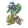
| ||||||||
|---|---|---|---|---|---|---|---|---|---|
| 1 |
| ||||||||
| Unit cell |
| ||||||||
| Details | The biological assembly is the heterotetramer present in the asymmetric unit |
-Components
-Protein , 2 types, 4 molecules ACBD
| #1: Protein | Mass: 14830.991 Da / Num. of mol.: 2 Source method: isolated from a genetically manipulated source Source: (gene. exp.) Clostridium cochlearium (bacteria) / Gene: GLMS / Plasmid: POZ3 / Production host: #2: Protein | Mass: 53623.172 Da / Num. of mol.: 2 Source method: isolated from a genetically manipulated source Details: COMPLEXED WITH ADENOSYLCOBALAMAIN AND A MIXTURE OF GLUTAMATE AND 2S,3S-3-METHYLASPARTIC ACID Source: (gene. exp.) Clostridium cochlearium (bacteria) / Gene: GLME / Plasmid: POZ5 / Production host: |
|---|
-Non-polymers , 5 types, 1266 molecules 


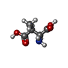

| #3: Chemical |  Mass: 1330.356 Da / Num. of mol.: 2 / Source method: obtained synthetically / Formula: C62H89CoN13O14P Mass: 1330.356 Da / Num. of mol.: 2 / Source method: obtained synthetically / Formula: C62H89CoN13O14P#4: Chemical |  Mass: 251.242 Da / Num. of mol.: 2 / Source method: obtained synthetically / Formula: C10H13N5O3 Mass: 251.242 Da / Num. of mol.: 2 / Source method: obtained synthetically / Formula: C10H13N5O3#5: Chemical |  Type: L-peptide linking / Mass: 147.129 Da / Num. of mol.: 2 / Source method: obtained synthetically / Formula: C5H9NO4 Type: L-peptide linking / Mass: 147.129 Da / Num. of mol.: 2 / Source method: obtained synthetically / Formula: C5H9NO4#6: Chemical |  Type: L-peptide linking / Mass: 147.129 Da / Num. of mol.: 2 / Source method: obtained synthetically / Formula: C5H9NO4 Type: L-peptide linking / Mass: 147.129 Da / Num. of mol.: 2 / Source method: obtained synthetically / Formula: C5H9NO4#7: Water | ChemComp-HOH / | Mass: 18.015 Da / Num. of mol.: 1258 / Source method: isolated from a natural source / Formula: H2O |
|---|
-Experimental details
-Experiment
| Experiment | Method: X-RAY DIFFRACTION / Number of used crystals: 1 |
|---|
- Sample preparation
| Crystal | Density Matthews: 2.4 Å3/Da / Density % sol: 48.7 % | ||||||||||||||||||||||||||||||||||||||||||
|---|---|---|---|---|---|---|---|---|---|---|---|---|---|---|---|---|---|---|---|---|---|---|---|---|---|---|---|---|---|---|---|---|---|---|---|---|---|---|---|---|---|---|---|
| Crystal grow | Temperature: 293 K / Method: vapor diffusion, sitting drop / pH: 4.6 Details: PEG-4000, cadmium chloride, coenzyme B12, sodium glutamate, pH 4.6, VAPOR DIFFUSION, SITTING DROP, temperature 293K | ||||||||||||||||||||||||||||||||||||||||||
| Crystal grow | *PLUS pH: 4.5 | ||||||||||||||||||||||||||||||||||||||||||
| Components of the solutions | *PLUS
|
-Data collection
| Diffraction | Mean temperature: 103 K |
|---|---|
| Diffraction source | Source: SYNCHROTRON / Site: EMBL/DESY, HAMBURG  / Beamline: BW7B / Wavelength: 0.8439 / Beamline: BW7B / Wavelength: 0.8439 |
| Detector | Type: MARRESEARCH / Detector: IMAGE PLATE / Date: Aug 15, 1999 |
| Radiation | Protocol: SINGLE WAVELENGTH / Monochromatic (M) / Laue (L): M / Scattering type: x-ray |
| Radiation wavelength | Wavelength: 0.8439 Å / Relative weight: 1 |
| Reflection | Resolution: 1.9→30 Å / Num. all: 121680 / Num. obs: 121680 / % possible obs: 99.9 % / Observed criterion σ(F): 0 / Observed criterion σ(I): 0 / Redundancy: 4.6 % / Biso Wilson estimate: 15.5 Å2 / Rmerge(I) obs: 0.073 / Rsym value: 0.073 / Net I/σ(I): 18.4 |
| Reflection shell | Resolution: 1.9→1.94 Å / Redundancy: 3.7 % / Rmerge(I) obs: 0.281 / Mean I/σ(I) obs: 5 / Num. unique all: 8102 / Rsym value: 0.281 / % possible all: 100 |
| Reflection | *PLUS Num. measured all: 555148 |
| Reflection shell | *PLUS % possible obs: 100 % / Num. unique obs: 8102 / Num. measured obs: 30037 |
- Processing
| Software |
| |||||||||||||||||||||||||||||||||
|---|---|---|---|---|---|---|---|---|---|---|---|---|---|---|---|---|---|---|---|---|---|---|---|---|---|---|---|---|---|---|---|---|---|---|
| Refinement | Method to determine structure: RIGID BODY REFINEMENT Starting model: 1CCW Resolution: 1.9→30 Å / Num. parameters: 45086 / Num. restraintsaints: 56174 / Isotropic thermal model: ISOTROPIC / Cross valid method: FREE R / σ(F): 0 / σ(I): 0 / Stereochemistry target values: ENGH AND HUBER Details: ANISOTROPIC SCALING APPLIED BY THE METHOD OF PARKIN, MOEZZI & HOPE, J.APPL.CRYST.28(1995)53-56
| |||||||||||||||||||||||||||||||||
| Solvent computation | Solvent model: MOEWS & KRETSINGER, J.MOL.BIOL.91(1973)201-228 | |||||||||||||||||||||||||||||||||
| Displacement parameters | Biso mean: 13.3 Å2 | |||||||||||||||||||||||||||||||||
| Refine analyze | Num. disordered residues: 39 / Occupancy sum hydrogen: 9804 / Occupancy sum non hydrogen: 11102 | |||||||||||||||||||||||||||||||||
| Refinement step | Cycle: LAST / Resolution: 1.9→30 Å
| |||||||||||||||||||||||||||||||||
| Refine LS restraints |
| |||||||||||||||||||||||||||||||||
| Software | *PLUS Name: SHELXL / Version: 97 / Classification: refinement | |||||||||||||||||||||||||||||||||
| Refinement | *PLUS σ(F): 0 / % reflection Rfree: 5 % / Rfactor obs: 0.168 | |||||||||||||||||||||||||||||||||
| Solvent computation | *PLUS | |||||||||||||||||||||||||||||||||
| Displacement parameters | *PLUS | |||||||||||||||||||||||||||||||||
| Refine LS restraints | *PLUS
|
