 Movie
Movie Controller

[English] 日本語
 Yorodumi
- PDB-6h9e: Structure of glutamate mutase reconstituted with homo-coenzyme B12 -
+ Open data

- Basic information
| Entry | Database: PDB / ID: 6h9e | ||||||
|---|---|---|---|---|---|---|---|
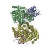
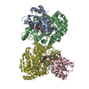
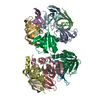
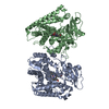
| Title | Structure of glutamate mutase reconstituted with homo-coenzyme B12 | ||||||
 Components Components | (Glutamate mutase ...) x 2 | ||||||
 Keywords Keywords | ISOMERASE / COENZYME B12 / CO-C-BOND / RADICAL REACTION / TIM-BARREL / ROSSMAN-FOLD | ||||||
| Function / homology |  Function and homology information Function and homology informationmethylaspartate mutase / : / methylaspartate mutase activity / : / cobalamin binding / metal ion binding Similarity search - Function | ||||||
| Biological species |  Clostridium cochlearium (bacteria) Clostridium cochlearium (bacteria) | ||||||
| Method |  X-RAY DIFFRACTION / SYNCHROTRON / MOLECULAR REPLACEMENT / Resolution: 1.82 Å X-RAY DIFFRACTION / SYNCHROTRON / MOLECULAR REPLACEMENT / Resolution: 1.82 Å | ||||||
 Authors Authors | Gruber, K. / Csitkovits, V. / Kratky, C. | ||||||
| Funding support |  Austria, 1items Austria, 1items
| ||||||
 Citation Citation | Journal: Angew.Chem.Int.Ed.Engl. / Year: 2022 Title: Structure-Based Demystification of Radical Catalysis by a Coenzyme B 12 Dependent Enzyme-Crystallographic Study of Glutamate Mutase with Cofactor Homologues. Authors: Gruber, K. / Csitkovits, V. / Lyskowski, A. / Kratky, C. / Krautler, B. | ||||||
| History |
|
- Structure visualization
| Structure viewer | Molecule: MolmilJmol/JSmol |
|---|
- Downloads & links
-Download
| PDBx/mmCIF format | 6h9e.cif.gz | 366.4 KB | Display | PDBx/mmCIF format |
|---|---|---|---|---|
| PDB format | pdb6h9e.ent.gz | 234.5 KB | Display | PDB format |
| PDBx/mmJSON format | 6h9e.json.gz | Tree view | PDBx/mmJSON format | |
| Others |  Other downloads Other downloads |
-Validation report
| Arichive directory | https://data.pdbj.org/pub/pdb/validation_reports/h9/6h9eftp://data.pdbj.org/pub/pdb/validation_reports/h9/6h9e | HTTPS FTP |
|---|
-Related structure data
| Related structure data |  6h9fC  1ccwS S: Starting model for refinement C: citing same article ( |
|---|---|
| Similar structure data |


-Links
 PDBj
PDBj
- Assembly
| Deposited unit | 
| ||||||||||||
|---|---|---|---|---|---|---|---|---|---|---|---|---|---|
| 1 |
| ||||||||||||
| Unit cell |
|
-Components
-Glutamate mutase ... , 2 types, 4 molecules ACBD
| #1: Protein | Mass: 14830.991 Da / Num. of mol.: 2 Source method: isolated from a genetically manipulated source Details: small subunit / Source: (gene. exp.) Clostridium cochlearium (bacteria) / Gene: glmS / Production host: #2: Protein | Mass: 53614.145 Da / Num. of mol.: 2 Source method: isolated from a genetically manipulated source Details: large subunit / Source: (gene. exp.) Clostridium cochlearium (bacteria) / Gene: glmE / Production host: |
|---|
-Non-polymers , 5 types, 1483 molecules 


| #3: Chemical |  Mass: 1330.356 Da / Num. of mol.: 2 / Source method: obtained synthetically / Formula: C62H89CoN13O14P / Feature type: SUBJECT OF INVESTIGATION Mass: 1330.356 Da / Num. of mol.: 2 / Source method: obtained synthetically / Formula: C62H89CoN13O14P / Feature type: SUBJECT OF INVESTIGATION#4: Chemical |  Mass: 265.269 Da / Num. of mol.: 2 / Source method: obtained synthetically / Formula: C11H15N5O3 / Feature type: SUBJECT OF INVESTIGATION Mass: 265.269 Da / Num. of mol.: 2 / Source method: obtained synthetically / Formula: C11H15N5O3 / Feature type: SUBJECT OF INVESTIGATION#5: Chemical |  Mass: 150.087 Da / Num. of mol.: 2 / Source method: obtained synthetically / Formula: C4H6O6 Mass: 150.087 Da / Num. of mol.: 2 / Source method: obtained synthetically / Formula: C4H6O6#6: Chemical | ChemComp-GOL /  Mass: 92.094 Da / Num. of mol.: 5 / Source method: obtained synthetically / Formula: C3H8O3 Mass: 92.094 Da / Num. of mol.: 5 / Source method: obtained synthetically / Formula: C3H8O3#7: Water | ChemComp-HOH / | Mass: 18.015 Da / Num. of mol.: 1472 / Source method: isolated from a natural source / Formula: H2O |
|---|
-Experimental details
-Experiment
| Experiment | Method: X-RAY DIFFRACTION / Number of used crystals: 1 |
|---|
- Sample preparation
| Crystal | Density Matthews: 2.83 Å3/Da / Density % sol: 56.59 % |
|---|---|
| Crystal grow | Temperature: 293 K / Method: vapor diffusion, sitting drop / pH: 4.5 Details: 6% (w/v) PEG-4000, 0.1 M DL-tartrate, pH=4.5, 2 mM CdCl2 |
-Data collection
| Diffraction | Mean temperature: 100 K |
|---|---|
| Diffraction source | Source: SYNCHROTRON / Site: EMBL/DESY, HAMBURG  / Beamline: BW7B / Wavelength: 0.8428 Å / Beamline: BW7B / Wavelength: 0.8428 Å |
| Detector | Type: MARRESEARCH / Detector: IMAGE PLATE / Date: Feb 5, 2000 |
| Radiation | Protocol: SINGLE WAVELENGTH / Monochromatic (M) / Laue (L): M / Scattering type: x-ray |
| Radiation wavelength | Wavelength: 0.8428 Å / Relative weight: 1 |
| Reflection | Resolution: 1.82→40.41 Å / Num. obs: 130639 / % possible obs: 96.1 % / Redundancy: 3 % / Biso Wilson estimate: 13.42 Å2 / CC1/2: 0.998 / Rmerge(I) obs: 0.0525 / Rpim(I) all: 0.0352 / Rrim(I) all: 0.0635 / Net I/σ(I): 19.5 |
| Reflection shell | Resolution: 1.82→1.89 Å / Redundancy: 2.8 % / Rmerge(I) obs: 0.2994 / Mean I/σ(I) obs: 4.5 / Num. unique obs: 12636 / CC1/2: 0.915 / Rpim(I) all: 0.2071 / Rrim(I) all: 0.3658 / % possible all: 93.7 |
- Processing
| Software |
| |||||||||||||||||||||||||||||||||||||||||||||||||||||||||||||||||||||||||||||||||||||||||||||||||||||||||||||||||||||||||||||||||||||||||||||||||||||||||||||||||||||||||||||||||||||||||||||||||||||||||||||||||||||||||
|---|---|---|---|---|---|---|---|---|---|---|---|---|---|---|---|---|---|---|---|---|---|---|---|---|---|---|---|---|---|---|---|---|---|---|---|---|---|---|---|---|---|---|---|---|---|---|---|---|---|---|---|---|---|---|---|---|---|---|---|---|---|---|---|---|---|---|---|---|---|---|---|---|---|---|---|---|---|---|---|---|---|---|---|---|---|---|---|---|---|---|---|---|---|---|---|---|---|---|---|---|---|---|---|---|---|---|---|---|---|---|---|---|---|---|---|---|---|---|---|---|---|---|---|---|---|---|---|---|---|---|---|---|---|---|---|---|---|---|---|---|---|---|---|---|---|---|---|---|---|---|---|---|---|---|---|---|---|---|---|---|---|---|---|---|---|---|---|---|---|---|---|---|---|---|---|---|---|---|---|---|---|---|---|---|---|---|---|---|---|---|---|---|---|---|---|---|---|---|---|---|---|---|---|---|---|---|---|---|---|---|---|---|---|---|---|---|---|---|
| Refinement | Method to determine structure: MOLECULAR REPLACEMENT Starting model: 1ccw Resolution: 1.82→40.41 Å / SU ML: 0.1594 / Cross valid method: FREE R-VALUE / σ(F): 1.34 / Phase error: 16.1663
| |||||||||||||||||||||||||||||||||||||||||||||||||||||||||||||||||||||||||||||||||||||||||||||||||||||||||||||||||||||||||||||||||||||||||||||||||||||||||||||||||||||||||||||||||||||||||||||||||||||||||||||||||||||||||
| Solvent computation | Shrinkage radii: 0.9 Å / VDW probe radii: 1.11 Å | |||||||||||||||||||||||||||||||||||||||||||||||||||||||||||||||||||||||||||||||||||||||||||||||||||||||||||||||||||||||||||||||||||||||||||||||||||||||||||||||||||||||||||||||||||||||||||||||||||||||||||||||||||||||||
| Displacement parameters | Biso mean: 14.92 Å2 | |||||||||||||||||||||||||||||||||||||||||||||||||||||||||||||||||||||||||||||||||||||||||||||||||||||||||||||||||||||||||||||||||||||||||||||||||||||||||||||||||||||||||||||||||||||||||||||||||||||||||||||||||||||||||
| Refinement step | Cycle: LAST / Resolution: 1.82→40.41 Å
| |||||||||||||||||||||||||||||||||||||||||||||||||||||||||||||||||||||||||||||||||||||||||||||||||||||||||||||||||||||||||||||||||||||||||||||||||||||||||||||||||||||||||||||||||||||||||||||||||||||||||||||||||||||||||
| Refine LS restraints |
| |||||||||||||||||||||||||||||||||||||||||||||||||||||||||||||||||||||||||||||||||||||||||||||||||||||||||||||||||||||||||||||||||||||||||||||||||||||||||||||||||||||||||||||||||||||||||||||||||||||||||||||||||||||||||
| LS refinement shell |
|
