[English] 日本語
Yorodumi
- PDB-2uvf: Structure of Yersinia enterocolitica Family 28 Exopolygalacturona... -

+
Open data


ID or keywords:

Loading...

-
Basic information

Entry
Database: PDB / ID: 2uvf
TitleStructure of Yersinia enterocolitica Family 28 Exopolygalacturonase in Complex with Digalaturonic Acid
ComponentsEXOPOLYGALACTURONASE
KeywordsHYDROLASE / GH28 / PECTIN / CELL WALL / PERIPLASM / YERSINIA ENTEROCOLITICA / BETA-HELIX / GLYCOSIDASE / EXO-ACTIVITY
Function / homology
Function and homology information


polygalacturonase activity / carbohydrate metabolic process
Similarity search - Function
Glycoside hydrolase, family 28 / Glycosyl hydrolases family 28 / Pectate lyase superfamily protein / Pectate lyase superfamily protein / Single-stranded right-handed beta-helix, Pectin lyase-like / Pectate Lyase C-like / Pectin lyase fold / Pectin lyase fold/virulence factor / 3 Solenoid / Fibronectin type 3 domain ...Glycoside hydrolase, family 28 / Glycosyl hydrolases family 28 / Pectate lyase superfamily protein / Pectate lyase superfamily protein / Single-stranded right-handed beta-helix, Pectin lyase-like / Pectate Lyase C-like / Pectin lyase fold / Pectin lyase fold/virulence factor / 3 Solenoid / Fibronectin type 3 domain / Fibronectin type-III domain profile. / Fibronectin type III / Fibronectin type III superfamily / Immunoglobulins / Immunoglobulin-like fold / Immunoglobulin-like / Sandwich / Mainly Beta
Similarity search - Domain/homology
ACETATE ION / NICKEL (II) ION / DI(HYDROXYETHYL)ETHER / Exopolygalacturonase
Similarity search - Component
Biological speciesYERSINIA ENTEROCOLITICA (bacteria)
MethodX-RAY DIFFRACTION / MOLECULAR REPLACEMENT / Resolution: 2.1 Å
AuthorsAbbott, D.W. / Boraston, A.B.
CitationJournal: J.Mol.Biol. / Year: 2007
Title: The Structural Basis for Exopolygalacturonase Activity in a Family 28 Glycoside Hydrolase.
Authors: Abbott, D.W. / Boraston, A.B.
History
DepositionMar 9, 2007Deposition site: PDBE / Processing site: PDBE
Revision 1.0May 8, 2007Provider: repository / Type: Initial release
Revision 1.1May 8, 2011Group: Version format compliance
Revision 1.2Jul 13, 2011Group: Version format compliance
Revision 2.0Jul 29, 2020Group: Advisory / Atomic model ...Advisory / Atomic model / Data collection / Derived calculations / Non-polymer description / Other / Structure summary
Category: atom_site / chem_comp ...atom_site / chem_comp / database_PDB_caveat / entity / pdbx_branch_scheme / pdbx_chem_comp_identifier / pdbx_database_status / pdbx_entity_branch / pdbx_entity_branch_descriptor / pdbx_entity_branch_link / pdbx_entity_branch_list / pdbx_entity_nonpoly / pdbx_nonpoly_scheme / pdbx_struct_conn_angle / struct_asym / struct_conn / struct_conn_type / struct_site / struct_site_gen
Item: _atom_site.B_iso_or_equiv / _atom_site.Cartn_x ..._atom_site.B_iso_or_equiv / _atom_site.Cartn_x / _atom_site.Cartn_y / _atom_site.Cartn_z / _atom_site.auth_asym_id / _atom_site.auth_atom_id / _atom_site.auth_comp_id / _atom_site.auth_seq_id / _atom_site.label_asym_id / _atom_site.label_atom_id / _atom_site.label_comp_id / _atom_site.label_entity_id / _atom_site.type_symbol / _chem_comp.formula / _chem_comp.formula_weight / _chem_comp.id / _chem_comp.name / _chem_comp.pdbx_synonyms / _chem_comp.type / _entity.formula_weight / _entity.pdbx_description / _entity.pdbx_number_of_molecules / _entity.src_method / _entity.type / _pdbx_database_status.status_code_sf / _pdbx_struct_conn_angle.ptnr1_auth_asym_id / _pdbx_struct_conn_angle.ptnr1_auth_comp_id / _pdbx_struct_conn_angle.ptnr1_auth_seq_id / _pdbx_struct_conn_angle.ptnr1_label_asym_id / _pdbx_struct_conn_angle.ptnr1_label_atom_id / _pdbx_struct_conn_angle.ptnr1_label_comp_id / _pdbx_struct_conn_angle.ptnr1_label_seq_id / _pdbx_struct_conn_angle.ptnr2_auth_seq_id / _pdbx_struct_conn_angle.ptnr2_label_asym_id / _pdbx_struct_conn_angle.ptnr3_auth_asym_id / _pdbx_struct_conn_angle.ptnr3_auth_comp_id / _pdbx_struct_conn_angle.ptnr3_auth_seq_id / _pdbx_struct_conn_angle.ptnr3_label_asym_id / _pdbx_struct_conn_angle.ptnr3_label_atom_id / _pdbx_struct_conn_angle.ptnr3_label_comp_id / _pdbx_struct_conn_angle.ptnr3_label_seq_id / _pdbx_struct_conn_angle.value / _struct_asym.entity_id
Description: Carbohydrate remediation / Provider: repository / Type: Remediation
Revision 2.1Dec 13, 2023Group: Data collection / Database references ...Data collection / Database references / Refinement description / Structure summary
Category: chem_comp / chem_comp_atom ...chem_comp / chem_comp_atom / chem_comp_bond / database_2 / pdbx_initial_refinement_model
Item: _chem_comp.pdbx_synonyms / _database_2.pdbx_DOI / _database_2.pdbx_database_accession
Remark 700 
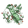
SHEET THE SHEET STRUCTURE OF THIS MOLECULE IS BIFURCATED. IN ORDER TO REPRESENT THIS FEATURE IN ... SHEET THE SHEET STRUCTURE OF THIS MOLECULE IS BIFURCATED. IN ORDER TO REPRESENT THIS FEATURE IN THE SHEET RECORDS BELOW, TWO SHEETS ARE DEFINED.

-
Structure visualization

Structure viewerMolecule:
MolmilJmol/JSmol

Downloads & links

-
Assembly

Deposited unit
A: EXOPOLYGALACTURONASE
B: EXOPOLYGALACTURONASE
hetero molecules


Theoretical massNumber of molelcules
Total (without water)136,59937
Polymers133,4642
Non-polymers3,13535
Water8,773487
1
A: EXOPOLYGALACTURONASE
hetero molecules


Theoretical massNumber of molelcules
Total (without water)67,91312
Polymers66,7321
Non-polymers1,18111
Water181
TypeNameSymmetry operationNumber
identity operation1_555x,y,z1
MethodPQS
2
B: EXOPOLYGALACTURONASE
hetero molecules


Theoretical massNumber of molelcules
Total (without water)68,68525
Polymers66,7321
Non-polymers1,95324
Water181
TypeNameSymmetry operationNumber
identity operation1_555x,y,z1
MethodPQS
Unit cell
Length a, b, c (Å)91.015, 79.659, 98.493
Angle α, β, γ (deg.)90.00, 103.90, 90.00
Int Tables number4
Space group name H-MP1211

-
Components

-
Protein / Sugars , 2 types, 3 molecules AB

#1: Protein EXOPOLYGALACTURONASE


Mass: 66731.984 Da / Num. of mol.: 2
Source method: isolated from a genetically manipulated source
Source: (gene. exp.) YERSINIA ENTEROCOLITICA (bacteria) / Production host: ESCHERICHIA COLI (E. coli) / References: UniProt: O68975*PLUS
#2: Polysaccharide alpha-D-galactopyranuronic acid-(1-4)-alpha-D-galactopyranuronic acid


Type: oligosaccharide / Mass: 370.263 Da / Num. of mol.: 1
Source method: isolated from a genetically manipulated source
DescriptorTypeProgram
DGalpAa1-4DGalpAa1-ROHGlycam Condensed SequenceGMML 1.0
WURCS=2.0/1,2,1/[a2112A-1a_1-5]/1-1/a4-b1WURCSPDB2Glycan 1.1.0
[][a-D-GalpA]{[(4+1)][a-D-GalpA]{}}LINUCSPDB-CARE

-
Non-polymers , 5 types, 521 molecules

#3: Chemical
ChemComp-SO4 / SULFATE ION / Sulfate


Mass: 96.063 Da / Num. of mol.: 18 / Source method: obtained synthetically / Formula: SO4
#4: Chemical
ChemComp-NI / NICKEL (II) ION / Nickel


Mass: 58.693 Da / Num. of mol.: 10 / Source method: obtained synthetically / Formula: Ni
#5: Chemical
ChemComp-ACT / ACETATE ION / Acetate


Mass: 59.044 Da / Num. of mol.: 4 / Source method: obtained synthetically / Formula: C2H3O2
#6: Chemical ChemComp-PEG / DI(HYDROXYETHYL)ETHER / Diethylene glycol


Mass: 106.120 Da / Num. of mol.: 2 / Source method: obtained synthetically / Formula: C4H10O3
#7: Water ChemComp-HOH / water / Water


Mass: 18.015 Da / Num. of mol.: 487 / Source method: isolated from a natural source / Formula: H2O

-
Details

Sequence detailsTHE AUTHORS REPORT THAT THIS IS MANUALLY SEQUENCED ENTRY FROM AN UNKNOWN STRAIN OF Y. ...THE AUTHORS REPORT THAT THIS IS MANUALLY SEQUENCED ENTRY FROM AN UNKNOWN STRAIN OF Y. ENTEROCOLITICA FROM THE ATCC STRAIN 9610. THIS STRAIN MATCHES THE SEQUENCED AND ANNOTATED ATCC 8081 STRAIN. THIS PARTICULAR SEQUENCE DOES NOT HAVE A UNIPROT ENTRY BUT DOES HAVE THE GENBANK CODE OF CAL10303.1.

-
Experimental details

-
Experiment

ExperimentMethod: X-RAY DIFFRACTION

-
Sample preparation

CrystalDensity Matthews: 2.61 Å3/Da / Density % sol: 52.59 %

-
Data collection

DiffractionMean temperature: 113 K
Diffraction sourceSource: ROTATING ANODE / Wavelength: 1.54
RadiationProtocol: SINGLE WAVELENGTH / Monochromatic (M) / Laue (L): M / Scattering type: x-ray
Radiation wavelengthWavelength: 1.54 Å / Relative weight: 1
ReflectionResolution: 2.1→19.72 Å / Num. obs: 75070 / % possible obs: 99.3 % / Observed criterion σ(I): 3 / Redundancy: 2.2 % / Rmerge(I) obs: 0.11

-
Processing

SoftwareName: REFMAC / Version: 5.2.0019 / Classification: refinement
RefinementMethod to determine structure: MOLECULAR REPLACEMENT
Starting model: PDB ENTRY 2UVE
Resolution: 2.1→19.72 Å / Cor.coef. Fo:Fc: 0.937 / Cor.coef. Fo:Fc free: 0.907 / SU B: 6.619 / SU ML: 0.176 / Cross valid method: THROUGHOUT / ESU R: 0.25 / ESU R Free: 0.216 / Stereochemistry target values: MAXIMUM LIKELIHOOD / Details: HYDROGENS HAVE BEEN ADDED IN THE RIDING POSITIONS
RfactorNum. reflection% reflectionSelection details
Rfree0.284 3976 5 %RANDOM
Rwork0.232 ---
obs0.234 75070 99.1 %-
Solvent computationIon probe radii: 0.8 Å / Shrinkage radii: 0.8 Å / VDW probe radii: 1.4 Å / Solvent model: MASK
Displacement parametersBiso mean: 34.58 Å2
Baniso -1Baniso -2Baniso -3
1--0.01 Å20 Å20 Å2
2--0.01 Å20 Å2
3---0 Å2
Refinement stepCycle: LAST / Resolution: 2.1→19.72 Å
ProteinNucleic acidLigandSolventTotal
Num. atoms8815 0 155 487 9457
Refine LS restraints
Refine-IDTypeDev idealDev ideal targetNumber
X-RAY DIFFRACTIONr_bond_refined_d0.0140.0229125
X-RAY DIFFRACTIONr_bond_other_d
X-RAY DIFFRACTIONr_angle_refined_deg1.5841.95712391
X-RAY DIFFRACTIONr_angle_other_deg
X-RAY DIFFRACTIONr_dihedral_angle_1_deg9.36651135
X-RAY DIFFRACTIONr_dihedral_angle_2_deg38.34125.255432
X-RAY DIFFRACTIONr_dihedral_angle_3_deg16.745151485
X-RAY DIFFRACTIONr_dihedral_angle_4_deg19.721542
X-RAY DIFFRACTIONr_chiral_restr0.1780.21356
X-RAY DIFFRACTIONr_gen_planes_refined0.0060.026966
X-RAY DIFFRACTIONr_gen_planes_other
X-RAY DIFFRACTIONr_nbd_refined0.2150.24335
X-RAY DIFFRACTIONr_nbd_other
X-RAY DIFFRACTIONr_nbtor_refined0.3070.26011
X-RAY DIFFRACTIONr_nbtor_other
X-RAY DIFFRACTIONr_xyhbond_nbd_refined0.1660.2601
X-RAY DIFFRACTIONr_xyhbond_nbd_other
X-RAY DIFFRACTIONr_metal_ion_refined
X-RAY DIFFRACTIONr_metal_ion_other
X-RAY DIFFRACTIONr_symmetry_vdw_refined0.2340.253
X-RAY DIFFRACTIONr_symmetry_vdw_other
X-RAY DIFFRACTIONr_symmetry_hbond_refined0.2230.26
X-RAY DIFFRACTIONr_symmetry_hbond_other
X-RAY DIFFRACTIONr_symmetry_metal_ion_refined
X-RAY DIFFRACTIONr_symmetry_metal_ion_other
X-RAY DIFFRACTIONr_mcbond_it0.7661.55814
X-RAY DIFFRACTIONr_mcbond_other
X-RAY DIFFRACTIONr_mcangle_it1.29929117
X-RAY DIFFRACTIONr_mcangle_other
X-RAY DIFFRACTIONr_scbond_it1.85633746
X-RAY DIFFRACTIONr_scbond_other
X-RAY DIFFRACTIONr_scangle_it2.864.53274
X-RAY DIFFRACTIONr_scangle_other
X-RAY DIFFRACTIONr_long_range_B_refined
X-RAY DIFFRACTIONr_long_range_B_other
X-RAY DIFFRACTIONr_rigid_bond_restr
X-RAY DIFFRACTIONr_sphericity_free
X-RAY DIFFRACTIONr_sphericity_bonded
LS refinement shellResolution: 2.1→2.15 Å / Total num. of bins used: 20
RfactorNum. reflection% reflection
Rfree0.391 301 -
Rwork0.324 5436 -
obs--98.74 %

+
About Yorodumi

-
News

-
Feb 9, 2022. New format data for meta-information of EMDB entries

New format data for meta-information of EMDB entries

  • Version 3 of the EMDB header file is now the official format.
  • The previous official version 1.9 will be removed from the archive.

Related info.:EMDB header

External links:wwPDB to switch to version 3 of the EMDB data model

-
Aug 12, 2020. Covid-19 info

Covid-19 info

URL: https://pdbj.org/emnavi/covid19.php

New page: Covid-19 featured information page in EM Navigator.

Related info.:Covid-19 info / Mar 5, 2020. Novel coronavirus structure data

+
Mar 5, 2020. Novel coronavirus structure data

Novel coronavirus structure data

Related info.:Yorodumi Speices / Aug 12, 2020. Covid-19 info

External links:COVID-19 featured content - PDBj / Molecule of the Month (242):Coronavirus Proteases

+
Jan 31, 2019. EMDB accession codes are about to change! (news from PDBe EMDB page)

EMDB accession codes are about to change! (news from PDBe EMDB page)

  • The allocation of 4 digits for EMDB accession codes will soon come to an end. Whilst these codes will remain in use, new EMDB accession codes will include an additional digit and will expand incrementally as the available range of codes is exhausted. The current 4-digit format prefixed with “EMD-” (i.e. EMD-XXXX) will advance to a 5-digit format (i.e. EMD-XXXXX), and so on. It is currently estimated that the 4-digit codes will be depleted around Spring 2019, at which point the 5-digit format will come into force.
  • The EM Navigator/Yorodumi systems omit the EMD- prefix.

Related info.:Q: What is EMD? / ID/Accession-code notation in Yorodumi/EM Navigator

External links:EMDB Accession Codes are Changing Soon! / Contact to PDBj

+
Jul 12, 2017. Major update of PDB

Major update of PDB

  • wwPDB released updated PDB data conforming to the new PDBx/mmCIF dictionary.
  • This is a major update changing the version number from 4 to 5, and with Remediation, in which all the entries are updated.
  • In this update, many items about electron microscopy experimental information are reorganized (e.g. em_software).
  • Now, EM Navigator and Yorodumi are based on the updated data.

External links:wwPDB Remediation / Enriched Model Files Conforming to OneDep Data Standards Now Available in the PDB FTP Archive

-
Yorodumi

Thousand views of thousand structures

  • Yorodumi is a browser for structure data from EMDB, PDB, SASBDB, etc.
  • This page is also the successor to EM Navigator detail page, and also detail information page/front-end page for Omokage search.
  • The word "yorodu" (or yorozu) is an old Japanese word meaning "ten thousand". "mi" (miru) is to see.

Related info.:EMDB / PDB / SASBDB / Comparison of 3 databanks / Yorodumi Search / Aug 31, 2016. New EM Navigator & Yorodumi / Yorodumi Papers / Jmol/JSmol / Function and homology information / Changes in new EM Navigator and Yorodumi

Read more