 Movie
Movie Controller

[English] 日本語
 Yorodumi
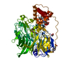
- PDB-7ae2: Crystal structure of HEPN(H107A-Y109F) toxin in complex with MNT ... -
+ Open data

- Basic information
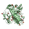
| Entry | Database: PDB / ID: 7ae2 | ||||||
|---|---|---|---|---|---|---|---|
| Title | Crystal structure of HEPN(H107A-Y109F) toxin in complex with MNT antitoxin | ||||||
 Components Components |
| ||||||
 Keywords Keywords | TOXIN/ANTITOXIN / toxin-antitoxin system / MNT-HEPN / AMPylation / TOXIN-ANTITOXIN complex | ||||||
| Function / homology |  Function and homology information Function and homology informationAMPylase activity / protein adenylyltransferase / toxin-antitoxin complex / Hydrolases; Acting on ester bonds; Endoribonucleases producing 3'-phosphomonoesters / RNA nuclease activity / endonuclease activity / nucleotide binding / hydrolase activity / ATP binding / metal ion binding Similarity search - Function | ||||||
| Biological species |  Aphanizomenon flos-aquae 2012/KM1/D3 (bacteria) Aphanizomenon flos-aquae 2012/KM1/D3 (bacteria) | ||||||
| Method |  X-RAY DIFFRACTION / SYNCHROTRON / SAD / Resolution: 2 Å X-RAY DIFFRACTION / SYNCHROTRON / SAD / Resolution: 2 Å | ||||||
 Authors Authors | Tamulaitiene, G. / Sasnauskas, G. / Songailiene, I. / Juozapaitis, J. / Siksnys, V. | ||||||
 Citation Citation | Journal: Mol.Cell / Year: 2020 Title: HEPN-MNT Toxin-Antitoxin System: The HEPN Ribonuclease Is Neutralized by OligoAMPylation. Authors: Songailiene, I. / Juozapaitis, J. / Tamulaitiene, G. / Ruksenaite, A. / Sulcius, S. / Sasnauskas, G. / Venclovas, C. / Siksnys, V. | ||||||
| History |
|
- Structure visualization
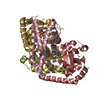
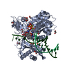
| Structure viewer | Molecule: MolmilJmol/JSmol |
|---|
- Downloads & links
-Download
| PDBx/mmCIF format | 7ae2.cif.gz | 129.3 KB | Display | PDBx/mmCIF format |
|---|---|---|---|---|
| PDB format | pdb7ae2.ent.gz | 100.1 KB | Display | PDB format |
| PDBx/mmJSON format | 7ae2.json.gz | Tree view | PDBx/mmJSON format | |
| Others |  Other downloads Other downloads |
-Validation report
| Arichive directory | https://data.pdbj.org/pub/pdb/validation_reports/ae/7ae2ftp://data.pdbj.org/pub/pdb/validation_reports/ae/7ae2 | HTTPS FTP |
|---|
-Related structure data


-Links
 PDBj
PDBj- Assembly
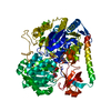
| Deposited unit | 
| ||||||||
|---|---|---|---|---|---|---|---|---|---|
| 1 | 
| ||||||||
| Unit cell |
| ||||||||
| Components on special symmetry positions |
|
-Components
| #1: Protein | Mass: 18257.652 Da / Num. of mol.: 1 / Mutation: H107A Y109F Source method: isolated from a genetically manipulated source Details: C-terminal His-tag Source: (gene. exp.) Aphanizomenon flos-aquae 2012/KM1/D3 (bacteria)Gene: OA07_26455 / Plasmid: pACYC-Duet / Production host: |
|---|---|
| #2: Protein | Mass: 17380.029 Da / Num. of mol.: 1 Source method: isolated from a genetically manipulated source Source: (gene. exp.) Aphanizomenon flos-aquae 2012/KM1/D3 (bacteria)Gene: OA07_26450 / Plasmid: pACYC-Duet / Production host: |
| #3: Water | ChemComp-HOH /  Mass: 18.015 Da / Num. of mol.: 66 / Source method: isolated from a natural source / Formula: H2O Mass: 18.015 Da / Num. of mol.: 66 / Source method: isolated from a natural source / Formula: H2O |
-Experimental details
-Experiment
| Experiment | Method: X-RAY DIFFRACTION / Number of used crystals: 1 |
|---|
- Sample preparation
| Crystal | Density Matthews: 2.2 Å3/Da / Density % sol: 47 % |
|---|---|
| Crystal grow | Temperature: 291 K / Method: vapor diffusion, sitting drop / pH: 6.5 Details: Crystallization buffer was 0,1 M MES-imidazole pH 6.5, 10% PEG 20K, 20% PEG550MME, 30 mM NaNO3, 30 mM Na2HPO4, 30 mM(NH4)2SO4 |
-Data collection
| Diffraction |
| |||||||||||||||||||||||||||||||||||||||||||||||||||||||||||||||||||||||||||||||||||||||||||||||||||||||||||||||||||||||||
|---|---|---|---|---|---|---|---|---|---|---|---|---|---|---|---|---|---|---|---|---|---|---|---|---|---|---|---|---|---|---|---|---|---|---|---|---|---|---|---|---|---|---|---|---|---|---|---|---|---|---|---|---|---|---|---|---|---|---|---|---|---|---|---|---|---|---|---|---|---|---|---|---|---|---|---|---|---|---|---|---|---|---|---|---|---|---|---|---|---|---|---|---|---|---|---|---|---|---|---|---|---|---|---|---|---|---|---|---|---|---|---|---|---|---|---|---|---|---|---|---|---|---|
| Diffraction source |
| |||||||||||||||||||||||||||||||||||||||||||||||||||||||||||||||||||||||||||||||||||||||||||||||||||||||||||||||||||||||||
| Detector |
| |||||||||||||||||||||||||||||||||||||||||||||||||||||||||||||||||||||||||||||||||||||||||||||||||||||||||||||||||||||||||
| Radiation |
| |||||||||||||||||||||||||||||||||||||||||||||||||||||||||||||||||||||||||||||||||||||||||||||||||||||||||||||||||||||||||
| Radiation wavelength |
| |||||||||||||||||||||||||||||||||||||||||||||||||||||||||||||||||||||||||||||||||||||||||||||||||||||||||||||||||||||||||
| Reflection | Resolution: 2→55.521 Å / Num. all: 21539 / Num. obs: 21539 / % possible obs: 99.9 % / Redundancy: 6.4 % / Biso Wilson estimate: 41.17 Å2 / Rpim(I) all: 0.019 / Rrim(I) all: 0.049 / Rsym value: 0.041 / Net I/av σ(I): 8.5 / Net I/σ(I): 22.3 / Num. measured all: 136995 | |||||||||||||||||||||||||||||||||||||||||||||||||||||||||||||||||||||||||||||||||||||||||||||||||||||||||||||||||||||||||
| Reflection shell | Diffraction-ID: 1
|

-Phasing
| Phasing | Method: SAD |
|---|
- Processing
| Software |
| ||||||||||||||||||||||||||||||||||||||||||||||||||||||||||||||||||||||||||||||||||||||||||||||||||||||||||||||||||||||||||||||||||||||||||||||||||||||||||||||||||||||||||||||||||||
|---|---|---|---|---|---|---|---|---|---|---|---|---|---|---|---|---|---|---|---|---|---|---|---|---|---|---|---|---|---|---|---|---|---|---|---|---|---|---|---|---|---|---|---|---|---|---|---|---|---|---|---|---|---|---|---|---|---|---|---|---|---|---|---|---|---|---|---|---|---|---|---|---|---|---|---|---|---|---|---|---|---|---|---|---|---|---|---|---|---|---|---|---|---|---|---|---|---|---|---|---|---|---|---|---|---|---|---|---|---|---|---|---|---|---|---|---|---|---|---|---|---|---|---|---|---|---|---|---|---|---|---|---|---|---|---|---|---|---|---|---|---|---|---|---|---|---|---|---|---|---|---|---|---|---|---|---|---|---|---|---|---|---|---|---|---|---|---|---|---|---|---|---|---|---|---|---|---|---|---|---|---|
| Refinement | Method to determine structure: SAD / Resolution: 2→50.053 Å / SU ML: 0.24 / Cross valid method: THROUGHOUT / σ(F): 0.26 / Phase error: 24.07 / Stereochemistry target values: ML Details: Dataset 1 (17-MAY-18) is the primary dataset used for refinement. Dataset 2 (24-NOV-18), Se-Met SAD, was used for phasing.
| ||||||||||||||||||||||||||||||||||||||||||||||||||||||||||||||||||||||||||||||||||||||||||||||||||||||||||||||||||||||||||||||||||||||||||||||||||||||||||||||||||||||||||||||||||||
| Solvent computation | Shrinkage radii: 0.9 Å / VDW probe radii: 1.11 Å / Solvent model: FLAT BULK SOLVENT MODEL | ||||||||||||||||||||||||||||||||||||||||||||||||||||||||||||||||||||||||||||||||||||||||||||||||||||||||||||||||||||||||||||||||||||||||||||||||||||||||||||||||||||||||||||||||||||
| Displacement parameters | Biso max: 133.55 Å2 / Biso mean: 58.0177 Å2 / Biso min: 21.78 Å2 | ||||||||||||||||||||||||||||||||||||||||||||||||||||||||||||||||||||||||||||||||||||||||||||||||||||||||||||||||||||||||||||||||||||||||||||||||||||||||||||||||||||||||||||||||||||
| Refinement step | Cycle: final / Resolution: 2→50.053 Å
| ||||||||||||||||||||||||||||||||||||||||||||||||||||||||||||||||||||||||||||||||||||||||||||||||||||||||||||||||||||||||||||||||||||||||||||||||||||||||||||||||||||||||||||||||||||
| Refine LS restraints |
| ||||||||||||||||||||||||||||||||||||||||||||||||||||||||||||||||||||||||||||||||||||||||||||||||||||||||||||||||||||||||||||||||||||||||||||||||||||||||||||||||||||||||||||||||||||
| LS refinement shell | Refine-ID: X-RAY DIFFRACTION / Rfactor Rfree error: 0
| ||||||||||||||||||||||||||||||||||||||||||||||||||||||||||||||||||||||||||||||||||||||||||||||||||||||||||||||||||||||||||||||||||||||||||||||||||||||||||||||||||||||||||||||||||||
| Refinement TLS params. | Method: refined / Refine-ID: X-RAY DIFFRACTION
| ||||||||||||||||||||||||||||||||||||||||||||||||||||||||||||||||||||||||||||||||||||||||||||||||||||||||||||||||||||||||||||||||||||||||||||||||||||||||||||||||||||||||||||||||||||
| Refinement TLS group |
|
