 Movie
Movie Controller

[English] 日本語
 Yorodumi
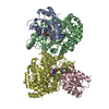
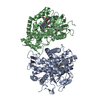
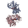
- PDB-1cb7: GLUTAMATE MUTASE FROM CLOSTRIDIUM COCHLEARIUM RECONSTITUTED WITH ... -
+ Open data

- Basic information
| Entry | Database: PDB / ID: 1cb7 | ||||||
|---|---|---|---|---|---|---|---|
| Title | GLUTAMATE MUTASE FROM CLOSTRIDIUM COCHLEARIUM RECONSTITUTED WITH METHYL-COBALAMIN | ||||||
 Components Components | (PROTEIN (GLUTAMATE MUTASE)) x 2 | ||||||
 Keywords Keywords | ISOMERASE / GLUTAMATE MUTASE / COENZYME-B12 / RADICAL REACTION / TIM-BARREL / ROSSMAN-FOLD | ||||||
| Function / homology |  Function and homology information Function and homology informationmethylaspartate mutase / : / methylaspartate mutase activity / : / cobalamin binding / metal ion binding Similarity search - Function | ||||||
| Biological species |  Clostridium cochlearium (bacteria) Clostridium cochlearium (bacteria) | ||||||
| Method |  X-RAY DIFFRACTION / SYNCHROTRON / MOLECULAR REPLACEMENT / Resolution: 2 Å X-RAY DIFFRACTION / SYNCHROTRON / MOLECULAR REPLACEMENT / Resolution: 2 Å | ||||||
 Authors Authors | Gruber, K. / Reitzer, R. / Kratky, C. | ||||||
 Citation Citation | Journal: Structure Fold.Des. / Year: 1999 Title: Glutamate mutase from Clostridium cochlearium: the structure of a coenzyme B12-dependent enzyme provides new mechanistic insights. Authors: Reitzer, R. / Gruber, K. / Jogl, G. / Wagner, U.G. / Bothe, H. / Buckel, W. / Kratky, C. #1: Journal: Acta Crystallogr.,Sect.D / Year: 1998 Title: Crystallization and preliminary X-ray analysis of recombinant glutamate mutase and of the isolated component S from Clostridium cochlearium. Authors: Reitzer, R. / Krasser, M. / Jogl, G. / Buckel, W. / Bothe, H. / Kratky, C. #2: Journal: Eur.J.Biochem. / Year: 1994 Title: Characterization of the coenzyme-B12-dependent glutamate mutase from Clostridium cochlearium produced in Escherichia coli. Authors: Zelder, O. / Beatrix, B. / Leutbecher, U. / Buckel, W. | ||||||
| History |
|
- Structure visualization
| Structure viewer | Molecule: MolmilJmol/JSmol |
|---|
- Downloads & links
-Download
| PDBx/mmCIF format | 1cb7.cif.gz | 277.2 KB | Display | PDBx/mmCIF format |
|---|---|---|---|---|
| PDB format | pdb1cb7.ent.gz | 220.1 KB | Display | PDB format |
| PDBx/mmJSON format | 1cb7.json.gz | Tree view | PDBx/mmJSON format | |
| Others |  Other downloads Other downloads |
-Validation report
| Arichive directory | https://data.pdbj.org/pub/pdb/validation_reports/cb/1cb7ftp://data.pdbj.org/pub/pdb/validation_reports/cb/1cb7 | HTTPS FTP |
|---|
-Related structure data


-Links
 PDBj
PDBj
- Assembly
| Deposited unit | 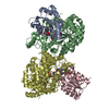
| ||||||||||
|---|---|---|---|---|---|---|---|---|---|---|---|
| 1 |
| ||||||||||
| Unit cell |
| ||||||||||
| Noncrystallographic symmetry (NCS) | NCS oper: (Code: given Matrix: (0.9804, -0.0042, 0.1969), Vector: |
-Components
| #1: Protein | Mass: 14830.046 Da / Num. of mol.: 2 Source method: isolated from a genetically manipulated source Details: CHAINS A, C, B, D FORM HETEROTETRAMER WHICH IS THE BIOLOGICAL UNIT. Source: (gene. exp.) Clostridium cochlearium (bacteria) / Gene: GLMS / Plasmid: POZ3 / Gene (production host): GLMS / Production host: #2: Protein | Mass: 53623.172 Da / Num. of mol.: 2 Source method: isolated from a genetically manipulated source Details: COMPLEXED WITH CO-METHYLCOBALAMIN / Source: (gene. exp.) Clostridium cochlearium (bacteria) / Gene: GLME / Plasmid: POZ5 / Gene (production host): GLME / Production host: #3: Chemical |   Mass: 1344.382 Da / Num. of mol.: 2 / Source method: obtained synthetically / Formula: C63H91CoN13O14P Mass: 1344.382 Da / Num. of mol.: 2 / Source method: obtained synthetically / Formula: C63H91CoN13O14P#4: Chemical |   Mass: 150.087 Da / Num. of mol.: 2 / Source method: obtained synthetically / Formula: C4H6O6 Mass: 150.087 Da / Num. of mol.: 2 / Source method: obtained synthetically / Formula: C4H6O6#5: Water | ChemComp-HOH / |  Mass: 18.015 Da / Num. of mol.: 1013 / Source method: isolated from a natural source / Formula: H2O Mass: 18.015 Da / Num. of mol.: 1013 / Source method: isolated from a natural source / Formula: H2O |
|---|
-Experimental details
-Experiment
| Experiment | Method: X-RAY DIFFRACTION / Number of used crystals: 1 |
|---|
- Sample preparation
| Crystal | Density Matthews: 2.87 Å3/Da / Density % sol: 57.19 % | ||||||||||||||||||||||||||||||||||||||||||||||||
|---|---|---|---|---|---|---|---|---|---|---|---|---|---|---|---|---|---|---|---|---|---|---|---|---|---|---|---|---|---|---|---|---|---|---|---|---|---|---|---|---|---|---|---|---|---|---|---|---|---|
| Crystal grow | pH: 4.5 / Details: pH 4.5 | ||||||||||||||||||||||||||||||||||||||||||||||||
| Components of the solutions |
| ||||||||||||||||||||||||||||||||||||||||||||||||
| Crystal grow | *PLUS pH: 7.5 / Method: vapor diffusion, hanging drop | ||||||||||||||||||||||||||||||||||||||||||||||||
| Components of the solutions | *PLUS
|
-Data collection
| Diffraction | Mean temperature: 105 K |
|---|---|
| Diffraction source | Source: SYNCHROTRON / Site: EMBL/DESY, HAMBURG  / Beamline: X11 / Wavelength: 0.9076 / Beamline: X11 / Wavelength: 0.9076 |
| Detector | Type: MARRESEARCH / Detector: IMAGE PLATE / Date: Oct 1, 1998 |
| Radiation | Protocol: SINGLE WAVELENGTH / Monochromatic (M) / Laue (L): M / Scattering type: x-ray |
| Radiation wavelength | Wavelength: 0.9076 Å / Relative weight: 1 |
| Reflection | Resolution: 2→25 Å / Num. obs: 100016 / % possible obs: 96.1 % / Redundancy: 2.6 % / Rsym value: 0.076 / Net I/σ(I): 5.6 |
| Reflection shell | Resolution: 2→2.11 Å / Redundancy: 2.5 % / Mean I/σ(I) obs: 1.3 / Rsym value: 0.189 / % possible all: 96.2 |
| Reflection | *PLUS Num. measured all: 258385 / Rmerge(I) obs: 0.076 |
| Reflection shell | *PLUS % possible obs: 96.2 % / Rmerge(I) obs: 0.189 |
- Processing
| Software |
| |||||||||||||||||||||||||||||||||
|---|---|---|---|---|---|---|---|---|---|---|---|---|---|---|---|---|---|---|---|---|---|---|---|---|---|---|---|---|---|---|---|---|---|---|
| Refinement | Method to determine structure: MOLECULAR REPLACEMENT Starting model: GLUTAMATE MUTASE RECONSTITUTED WITH CYANOCOBALAMIN Resolution: 2→25 Å / Num. parameters: 43322 / Num. restraintsaints: 54994 / Cross valid method: FREE R / σ(F): 0 / Stereochemistry target values: ENGH AND HUBER Details: ANISOTROPIC SCALING APPLIED BY THE METHOD OF PARKIN, MOEZZI & HOPE, J.APPL.CRYST.28 (1995)53-56. ANISOTROPIC REFINEMENT REDUCED FREE R (NO CUTOFF) BY APPROX. 0.02
| |||||||||||||||||||||||||||||||||
| Solvent computation | Solvent model: MOEWS & KRETSINGER, J.MOL.BIOL.91(1973)201-203 | |||||||||||||||||||||||||||||||||
| Refine analyze | Num. disordered residues: 0 / Occupancy sum hydrogen: 9832 / Occupancy sum non hydrogen: 10823 | |||||||||||||||||||||||||||||||||
| Refinement step | Cycle: LAST / Resolution: 2→25 Å
| |||||||||||||||||||||||||||||||||
| Refine LS restraints |
| |||||||||||||||||||||||||||||||||
| Software | *PLUS Name: SHELXL-97 / Classification: refinement | |||||||||||||||||||||||||||||||||
| Refinement | *PLUS σ(F): 0 / % reflection Rfree: 10 % / Rfactor all: 0.166 / Rfactor Rwork: 0.16 | |||||||||||||||||||||||||||||||||
| Solvent computation | *PLUS | |||||||||||||||||||||||||||||||||
| Displacement parameters | *PLUS | |||||||||||||||||||||||||||||||||
| Refine LS restraints | *PLUS
|
