 Movie
Movie Controller

[English] 日本語
 Yorodumi
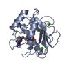
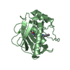
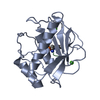
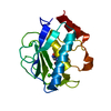
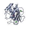
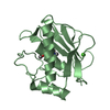
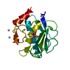
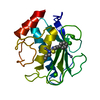
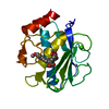
- PDB-1i76: COMPLEX OF 2-(BIPHENYL-4-SULFONYL)-1,2,3,4-TETRAHYDRO-ISOQUINOLIN... -
+ Open data

- Basic information
| Entry | Database: PDB / ID: 1i76 | ||||||
|---|---|---|---|---|---|---|---|
| Title | COMPLEX OF 2-(BIPHENYL-4-SULFONYL)-1,2,3,4-TETRAHYDRO-ISOQUINOLINE-3-CARBOXYLIC ACID (D-TIC DERIVATIVE) WITH T CATALITIC DOMAIN OF MATRIX METALLO PROTEINASE-8 (MET80 FORM) | ||||||
 Components Components | NEUTROPHIL COLLAGENASE | ||||||
 Keywords Keywords | HYDROLASE / COMPLEX (METALLOPROTEASE-INHIBITOR) | ||||||
| Function / homology |  Function and homology information Function and homology informationneutrophil collagenase / tumor necrosis factor binding / positive regulation of microglial cell activation / positive regulation of tumor necrosis factor-mediated signaling pathway / positive regulation of neuroinflammatory response / endodermal cell differentiation / Activation of Matrix Metalloproteinases / Collagen degradation / collagen catabolic process / extracellular matrix disassembly ...neutrophil collagenase / tumor necrosis factor binding / positive regulation of microglial cell activation / positive regulation of tumor necrosis factor-mediated signaling pathway / positive regulation of neuroinflammatory response / endodermal cell differentiation / Activation of Matrix Metalloproteinases / Collagen degradation / collagen catabolic process / extracellular matrix disassembly / Degradation of the extracellular matrix / extracellular matrix organization / metalloendopeptidase activity / specific granule lumen / tertiary granule lumen / positive regulation of tumor necrosis factor production / peptidase activity / cellular response to lipopolysaccharide / extracellular matrix / endopeptidase activity / serine-type endopeptidase activity / Neutrophil degranulation / proteolysis / : / extracellular region / zinc ion binding Similarity search - Function | ||||||
| Biological species |  Homo sapiens (human) Homo sapiens (human) | ||||||
| Method |  X-RAY DIFFRACTION / SYNCHROTRON / MOLECULAR REPLACEMENT / Resolution: 1.2 Å X-RAY DIFFRACTION / SYNCHROTRON / MOLECULAR REPLACEMENT / Resolution: 1.2 Å | ||||||
 Authors Authors | Gavuzzo, E. / Pochetti, G. / Mazza, F. / Gallina, C. / Gorini, B. / D'Alessio, S. / Pieper, M. / Tschesche, H. / Tucker, P.A. | ||||||
 Citation Citation | Journal: J.Med.Chem. / Year: 2000 Title: Two crystal structures of human neutrophil collagenase, one complexed with a primed- and the other with an unprimed-side inhibitor: implications for drug design. Authors: Gavuzzo, E. / Pochetti, G. / Mazza, F. / Gallina, C. / Gorini, B. / D'Alessio, S. / Pieper, M. / Tschesche, H. / Tucker, P.A. #1: Journal: J.Med.Chem. / Year: 1999Title: QUANTITATIVE STRUCTURE-ACTIVITY RELATIONSHIP OF HUMAN NEUTROPHIL COLLAGENASE (MMP8-8) INHIBITORS USING COMPARATIVE MOLECULAR FIELD ANALYSIS AND X-RAY STRUCTURE ANALYSIS Authors: Matter, H. / Schwab, W. / Barbier, D. / Billen, G. / Haase, B. / Schudok, M. / Neises, B. / Thorwart, W. / Schreuder, H. / Brachvogel, V. / Weithmann, K.U. / Loenze, P. #2: Journal: Eur.J.Biochem. / Year: 1995Title: X-RAY STRUCTURES OF HUMAN NEUTROPHIL COLLAGENASE COMPLEXED WITH PEPTIDE HYDROXAMATE AND PEPTIDE THIOL INHIBITORS. IMPLICATIONS FOR SUBSTRATE BINDING AND RATIONAL DRUG DESIGN Authors: Grams, F. / Reinemer, P. / Powers, J.C. / Kleine, T. / Pieper, M. / Tschesche, H. / Huber, R. / Bode, W. | ||||||
| History |
|
- Structure visualization
| Structure viewer | Molecule: MolmilJmol/JSmol |
|---|
- Downloads & links
-Download
| PDBx/mmCIF format | 1i76.cif.gz | 94.3 KB | Display | PDBx/mmCIF format |
|---|---|---|---|---|
| PDB format | pdb1i76.ent.gz | 69.7 KB | Display | PDB format |
| PDBx/mmJSON format | 1i76.json.gz | Tree view | PDBx/mmJSON format | |
| Others |  Other downloads Other downloads |
-Validation report
| Arichive directory | https://data.pdbj.org/pub/pdb/validation_reports/i7/1i76ftp://data.pdbj.org/pub/pdb/validation_reports/i7/1i76 | HTTPS FTP |
|---|
-Related structure data
| Related structure data |  1i73C  1japS S: Starting model for refinement C: citing same article ( |
|---|---|
| Similar structure data |


-Links
 PDBj
PDBj


- Assembly
| Deposited unit | 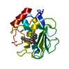
| ||||||||
|---|---|---|---|---|---|---|---|---|---|
| 1 |
| ||||||||
| Unit cell |
|
-Components
| #1: Protein | Mass: 18111.744 Da / Num. of mol.: 1 / Fragment: RESIDUES 80-242 Source method: isolated from a genetically manipulated source Source: (gene. exp.) Homo sapiens (human) / Production host:  | ||||||
|---|---|---|---|---|---|---|---|
| #2: Chemical |   Mass: 40.078 Da / Num. of mol.: 2 / Source method: obtained synthetically / Formula: Ca Mass: 40.078 Da / Num. of mol.: 2 / Source method: obtained synthetically / Formula: Ca#3: Chemical |   Mass: 65.409 Da / Num. of mol.: 2 / Source method: obtained synthetically / Formula: Zn Mass: 65.409 Da / Num. of mol.: 2 / Source method: obtained synthetically / Formula: Zn#4: Chemical | ChemComp-BSI / | 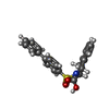  Mass: 393.456 Da / Num. of mol.: 1 / Source method: obtained synthetically / Formula: C22H19NO4S Mass: 393.456 Da / Num. of mol.: 1 / Source method: obtained synthetically / Formula: C22H19NO4S#5: Water | ChemComp-HOH / |  Mass: 18.015 Da / Num. of mol.: 271 / Source method: isolated from a natural source / Formula: H2O Mass: 18.015 Da / Num. of mol.: 271 / Source method: isolated from a natural source / Formula: H2O |
-Experimental details
-Experiment
| Experiment | Method: X-RAY DIFFRACTION / Number of used crystals: 1 |
|---|
- Sample preparation
| Crystal | Density Matthews: 2.21 Å3/Da / Density % sol: 44.25 % | ||||||||||||||||||||||||||||||||||||||||||||||||||||||||||||||||||||||
|---|---|---|---|---|---|---|---|---|---|---|---|---|---|---|---|---|---|---|---|---|---|---|---|---|---|---|---|---|---|---|---|---|---|---|---|---|---|---|---|---|---|---|---|---|---|---|---|---|---|---|---|---|---|---|---|---|---|---|---|---|---|---|---|---|---|---|---|---|---|---|---|
| Crystal grow | Temperature: 291 K / Method: vapor diffusion, hanging drop / pH: 6 Details: PEG 6000, MES-NAOH, NaCl, CaCl2, ZnCl2, pH 6.00, VAPOR DIFFUSION, HANGING DROP, temperature 291K | ||||||||||||||||||||||||||||||||||||||||||||||||||||||||||||||||||||||
| Crystal grow | *PLUS Temperature: 18 ℃Details: 2 micro litte of protein solution, 1 miro litter of inhibitor solution, and 5 micro litter of PEG solution | ||||||||||||||||||||||||||||||||||||||||||||||||||||||||||||||||||||||
| Components of the solutions | *PLUS
|
-Data collection
| Diffraction | Mean temperature: 100 K |
|---|---|
| Diffraction source | Source: SYNCHROTRON / Site: EMBL/DESY, HAMBURG  / Beamline: BW7B / Wavelength: 0.84 / Beamline: BW7B / Wavelength: 0.84 |
| Detector | Type: MAR scanner 345 mm plate / Detector: IMAGE PLATE / Date: Jul 21, 1998 |
| Radiation | Monochromator: MIRRORS / Protocol: SINGLE WAVELENGTH / Monochromatic (M) / Laue (L): M / Scattering type: x-ray |
| Radiation wavelength | Wavelength: 0.84 Å / Relative weight: 1 |
| Reflection | Resolution: 1.2→20 Å / Num. all: 51100 / Num. obs: 50709 / % possible obs: 99.3 % / Observed criterion σ(F): 0 / Observed criterion σ(I): 1 / Redundancy: 8.1 % / Biso Wilson estimate: 7.9 Å2 / Rmerge(I) obs: 0.09 / Net I/σ(I): 19.6 |
| Reflection shell | Resolution: 1.2→1.22 Å / Rmerge(I) obs: 0.177 / Mean I/σ(I) obs: 5 / % possible all: 95.3 |
| Reflection | *PLUS Num. measured all: 411807 |
| Reflection shell | *PLUS % possible obs: 95.3 % |
- Processing
| Software |
| |||||||||||||||||||||||||
|---|---|---|---|---|---|---|---|---|---|---|---|---|---|---|---|---|---|---|---|---|---|---|---|---|---|---|
| Refinement | Method to determine structure: MOLECULAR REPLACEMENT Starting model: PDB ENTRY 1JAP Resolution: 1.2→10 Å / Num. parameters: 14352 / Num. restraintsaints: 17304 / σ(F): 0 / σ(I): 0 / Stereochemistry target values: ENGH & HUBER
| |||||||||||||||||||||||||
| Refine analyze | Num. disordered residues: 5 / Occupancy sum hydrogen: 1201 / Occupancy sum non hydrogen: 1586 | |||||||||||||||||||||||||
| Refinement step | Cycle: LAST / Resolution: 1.2→10 Å
| |||||||||||||||||||||||||
| Refine LS restraints |
| |||||||||||||||||||||||||
| LS refinement shell | Resolution: 1.2→10 Å
|
