 Movie
Movie Controller

[English] 日本語
 Yorodumi
- PDB-1i35: SOLUTION STRUCTURE OF THE RAS-BINDING DOMAIN OF THE PROTEIN KINAS... -
+ Open data

- Basic information
| Entry | Database: PDB / ID: 1i35 | ||||||
|---|---|---|---|---|---|---|---|
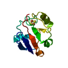
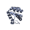
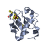
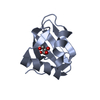
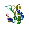
| Title | SOLUTION STRUCTURE OF THE RAS-BINDING DOMAIN OF THE PROTEIN KINASE BYR2 FROM SCHIZOSACCHAROMYCES POMBE | ||||||
 Components Components | PROTEIN KINASE BYR2 | ||||||
 Keywords Keywords | TRANSFERASE / Ubiquitin superfold | ||||||
| Function / homology |  Function and homology information Function and homology information: / pheromone response MAPK cascade / Oxidative Stress Induced Senescence / division septum / mitogen-activated protein kinase kinase kinase / cell tip / cell division site / p38MAPK cascade / MAP kinase kinase kinase activity / JNK cascade ...: / pheromone response MAPK cascade / Oxidative Stress Induced Senescence / division septum / mitogen-activated protein kinase kinase kinase / cell tip / cell division site / p38MAPK cascade / MAP kinase kinase kinase activity / JNK cascade / protein kinase activity / protein serine kinase activity / ATP binding / plasma membrane / cytoplasm Similarity search - Function | ||||||
| Biological species |  | ||||||
| Method | SOLUTION NMR / High temperature torsion angle dynamics. First cooling stage using torsion angle dynamics. Second cooling stage using Cartesian dynamics. Final energy minimization. | ||||||
 Authors Authors | Gronwald, W. / Huber, F. / Grunewald, P. / Sporner, M. / Wohlgemuth, S. / Herrmann, C. / Kalbitzer, H.R. | ||||||
 Citation Citation | Journal: Structure / Year: 2001 Title: Solution structure of the Ras binding domain of the protein kinase Byr2 from Schizosaccharomyces pombe. Authors: Gronwald, W. / Huber, F. / Grunewald, P. / Sporner, M. / Wohlgemuth, S. / Herrmann, C. / Kalbitzer, H.R. #1: Journal: Protein Sci. / Year: 2001Title: Overcoming the Problems Associated with Poor Spectra Quality of the Protein Kinase Byr2 using Residual Dipolar Couplings Authors: Gronwald, W. / Brunner, E. / Huber, F. / Wenzler, M. / Herrmann, C. / Kalbitzer, H.R. #2: Journal: J.Biomol.NMR / Year: 2000Title: Letter to the Editor: Sequential NMR assignment of the RAS-binding domain of Byr2 Authors: Huber, F. / Gronwald, W. / Wohlgemuth, S. / Herrmann, C. / Geyer, M. / Wittinghofer, A. / Kalbitzer, H.R. | ||||||
| History |
|
- Structure visualization
| Structure viewer | Molecule: MolmilJmol/JSmol |
|---|
- Downloads & links
-Download
| PDBx/mmCIF format | 1i35.cif.gz | 308.9 KB | Display | PDBx/mmCIF format |
|---|---|---|---|---|
| PDB format | pdb1i35.ent.gz | 254.9 KB | Display | PDB format |
| PDBx/mmJSON format | 1i35.json.gz | Tree view | PDBx/mmJSON format | |
| Others |  Other downloads Other downloads |
-Validation report
| Arichive directory | https://data.pdbj.org/pub/pdb/validation_reports/i3/1i35ftp://data.pdbj.org/pub/pdb/validation_reports/i3/1i35 | HTTPS FTP |
|---|
-Related structure data
| Similar structure data |
|---|


-Links
 PDBj
PDBj- Assembly
| Deposited unit | 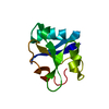
| |||||||||
|---|---|---|---|---|---|---|---|---|---|---|
| 1 |
| |||||||||
| NMR ensembles |
|
-Components
| #1: Protein | Mass: 10987.690 Da / Num. of mol.: 1 / Fragment: RAS-BINDING DOMAIN Source method: isolated from a genetically manipulated source Source: (gene. exp.) Gene: BYR2 (AMINO ACIDS 71 - 165) / Plasmid: PGEX4T3 (PHARMACIA) / Production host:  References: UniProt: P28829, Transferases; Transferring phosphorus-containing groups; Phosphotransferases with an alcohol group as acceptor |
|---|
-Experimental details
-Experiment
| Experiment | Method: SOLUTION NMR | ||||||||||||||||||||||||||||
|---|---|---|---|---|---|---|---|---|---|---|---|---|---|---|---|---|---|---|---|---|---|---|---|---|---|---|---|---|---|
| NMR experiment |
|
 HSQC
HSQC- Sample preparation
| Details |
| |||||||||||||||||||||||||||||||||||
|---|---|---|---|---|---|---|---|---|---|---|---|---|---|---|---|---|---|---|---|---|---|---|---|---|---|---|---|---|---|---|---|---|---|---|---|---|
| Sample conditions |
| |||||||||||||||||||||||||||||||||||
| Crystal grow | *PLUS Method: other / Details: NMR |
-NMR measurement
| Radiation | Protocol: SINGLE WAVELENGTH / Monochromatic (M) / Laue (L): M | |||||||||||||||
|---|---|---|---|---|---|---|---|---|---|---|---|---|---|---|---|---|
| Radiation wavelength | Relative weight: 1 | |||||||||||||||
| NMR spectrometer |
|
- Processing
| NMR software |
| ||||||||||||||||||||||||||||
|---|---|---|---|---|---|---|---|---|---|---|---|---|---|---|---|---|---|---|---|---|---|---|---|---|---|---|---|---|---|
| Refinement | Method: High temperature torsion angle dynamics. First cooling stage using torsion angle dynamics. Second cooling stage using Cartesian dynamics. Final energy minimization. Software ordinal: 1 Details: Structures are based on a total of 824 NOE-restraints, 88 backbone dihedral angle restrains, 29 hydrogen bonds and 28 residual dipolar couplings | ||||||||||||||||||||||||||||
| NMR representative | Selection criteria: lowest energy | ||||||||||||||||||||||||||||
| NMR ensemble | Conformer selection criteria: The submitted conformer models are the 10 structures with the lowest total energy Conformers calculated total number: 200 / Conformers submitted total number: 10 |
