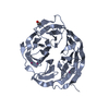
 Movie
Movie Controller


+ Open data

- Basic information
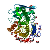
| Entry | Database: PDB / ID: 1h9s | ||||||
|---|---|---|---|---|---|---|---|
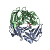
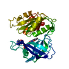
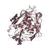
| Title | Molybdate bound complex of Dimop domain of ModE from E.coli | ||||||
 Components Components | (MOLYBDENUM TRANSPORT PROTEIN MODE) x 2 | ||||||
 Keywords Keywords | TRANSCRIPTION REGULATOR | ||||||
| Function / homology |  Function and homology information Function and homology informationModE complex / negative regulation of DNA-templated transcription initiation / molybdate ion transport / molybdenum ion binding / cis-regulatory region sequence-specific DNA binding / DNA-binding transcription factor activity / negative regulation of DNA-templated transcription / regulation of DNA-templated transcription / positive regulation of DNA-templated transcription / DNA binding ...ModE complex / negative regulation of DNA-templated transcription initiation / molybdate ion transport / molybdenum ion binding / cis-regulatory region sequence-specific DNA binding / DNA-binding transcription factor activity / negative regulation of DNA-templated transcription / regulation of DNA-templated transcription / positive regulation of DNA-templated transcription / DNA binding / cytoplasm / cytosol Similarity search - Function | ||||||
| Biological species |  | ||||||
| Method |  X-RAY DIFFRACTION / MOLECULAR REPLACEMENT / Resolution: 1.82 Å X-RAY DIFFRACTION / MOLECULAR REPLACEMENT / Resolution: 1.82 Å | ||||||
 Authors Authors | Gourley, D.G. / Hunter, W.N. | ||||||
 Citation Citation | Journal: J.Biol.Chem. / Year: 2001 Title: Oxyanion Binding Alters Conformational and Quaternary Structure of the C-Terminal Domain of the Transcriptional Regulator Mode; Implications for Molybdate-Dependant Regulation, Signalling, Storage and Transport Authors: Gourley, D.G. / Schuttelkopf, A.W. / Anderson, L.A. / Price, N.C. / Boxer, D.H. / Hunter, W.N. | ||||||
| History |
|
- Structure visualization
| Structure viewer | Molecule: MolmilJmol/JSmol |
|---|
- Downloads & links
-Download
| PDBx/mmCIF format | 1h9s.cif.gz | 69.3 KB | Display | PDBx/mmCIF format |
|---|---|---|---|---|
| PDB format | pdb1h9s.ent.gz | 50.4 KB | Display | PDB format |
| PDBx/mmJSON format | 1h9s.json.gz | Tree view | PDBx/mmJSON format | |
| Others |  Other downloads Other downloads |
-Validation report
| Arichive directory | https://data.pdbj.org/pub/pdb/validation_reports/h9/1h9sftp://data.pdbj.org/pub/pdb/validation_reports/h9/1h9s | HTTPS FTP |
|---|
-Related structure data
| Related structure data |  1h9rC  1b9nS S: Starting model for refinement C: citing same article ( |
|---|---|
| Similar structure data |


-Links
 PDBj
PDBj- Assembly
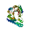
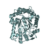
| Deposited unit | 
| ||||||||
|---|---|---|---|---|---|---|---|---|---|
| 1 |
| ||||||||
| Unit cell |
| ||||||||
| Noncrystallographic symmetry (NCS) | NCS oper: (Code: given Matrix: (0.125, -0.974, 0.187), Vector: |
-Components
| #1: Protein | Mass: 15048.901 Da / Num. of mol.: 1 / Fragment: MOLYBDATE BINDING DOMAIN RESIDUES 123-260 / Mutation: YES Source method: isolated from a genetically manipulated source Source: (gene. exp.) | ||||
|---|---|---|---|---|---|
| #2: Protein | Mass: 15049.888 Da / Num. of mol.: 1 / Fragment: MOLYBDATE BINDING DOMAIN RESIDUES 123-260 / Mutation: YES Source method: isolated from a genetically manipulated source Source: (gene. exp.) | ||||
| #3: Chemical |   Mass: 159.938 Da / Num. of mol.: 2 / Source method: obtained synthetically / Formula: MoO4 Mass: 159.938 Da / Num. of mol.: 2 / Source method: obtained synthetically / Formula: MoO4#4: Water | ChemComp-HOH / |  Mass: 18.015 Da / Num. of mol.: 227 / Source method: isolated from a natural source / Formula: H2O Mass: 18.015 Da / Num. of mol.: 227 / Source method: isolated from a natural source / Formula: H2OCompound details | CHAIN A, B ENGINEERED MUTATION FROM LEU 123 MET BINDS MOLYBDENUM. REGULATES MOD OPERON BY BINDING ...CHAIN A, B ENGINEERED | |
-Experimental details
-Experiment
| Experiment | Method: X-RAY DIFFRACTION / Number of used crystals: 1 |
|---|
- Sample preparation
| Crystal | Density Matthews: 2.18 Å3/Da / Density % sol: 43.68 % | ||||||||||||||||||||||||||||||||||||||||||||||||||||||||
|---|---|---|---|---|---|---|---|---|---|---|---|---|---|---|---|---|---|---|---|---|---|---|---|---|---|---|---|---|---|---|---|---|---|---|---|---|---|---|---|---|---|---|---|---|---|---|---|---|---|---|---|---|---|---|---|---|---|
| Crystal grow | pH: 7.6 / Details: pH 7.60 | ||||||||||||||||||||||||||||||||||||||||||||||||||||||||
| Crystal grow | *PLUS Temperature: 20 ℃ / Method: vapor diffusion, hanging drop | ||||||||||||||||||||||||||||||||||||||||||||||||||||||||
| Components of the solutions | *PLUS
|
-Data collection
| Diffraction | Mean temperature: 100 K |
|---|---|
| Diffraction source | Source: ROTATING ANODE / Type: RIGAKU RU200 / Wavelength: 1.5418 |
| Detector | Type: RIGAKU RAXIS IV / Detector: IMAGE PLATE / Details: MIRRORS |
| Radiation | Monochromator: NI FILTER / Protocol: SINGLE WAVELENGTH / Monochromatic (M) / Laue (L): M / Scattering type: x-ray |
| Radiation wavelength | Wavelength: 1.5418 Å / Relative weight: 1 |
| Reflection | Resolution: 1.9→30 Å / Num. obs: 20410 / % possible obs: 97.9 % / Redundancy: 9 % / Biso Wilson estimate: 27 Å2 / Rsym value: 0.49 / Net I/σ(I): 27.8 |
| Reflection | *PLUS Highest resolution: 1.82 Å / Lowest resolution: 30 Å / Num. obs: 22803 / % possible obs: 98.1 % / Num. measured all: 272924 / Rmerge(I) obs: 0.075 |
| Reflection shell | *PLUS % possible obs: 84.3 % / Rmerge(I) obs: 0.381 / Mean I/σ(I) obs: 2.9 |
- Processing
| Software |
| ||||||||||||||||||||||||||||||||||||||||||||||||||||||||||||||||||||||||||||||||||||
|---|---|---|---|---|---|---|---|---|---|---|---|---|---|---|---|---|---|---|---|---|---|---|---|---|---|---|---|---|---|---|---|---|---|---|---|---|---|---|---|---|---|---|---|---|---|---|---|---|---|---|---|---|---|---|---|---|---|---|---|---|---|---|---|---|---|---|---|---|---|---|---|---|---|---|---|---|---|---|---|---|---|---|---|---|---|
| Refinement | Method to determine structure: MOLECULAR REPLACEMENT Starting model: PDB ENTRY 1B9N Resolution: 1.82→20 Å / SU B: 2.95 / SU ML: 0.093 / Cross valid method: THROUGHOUT / σ(F): 0 / ESU R: 0.151 / ESU R Free: 0.144
| ||||||||||||||||||||||||||||||||||||||||||||||||||||||||||||||||||||||||||||||||||||
| Displacement parameters | Biso mean: 34.2 Å2 | ||||||||||||||||||||||||||||||||||||||||||||||||||||||||||||||||||||||||||||||||||||
| Refinement step | Cycle: LAST / Resolution: 1.82→20 Å
| ||||||||||||||||||||||||||||||||||||||||||||||||||||||||||||||||||||||||||||||||||||
| Refine LS restraints |
| ||||||||||||||||||||||||||||||||||||||||||||||||||||||||||||||||||||||||||||||||||||
| Software | *PLUS Name: REFMAC / Classification: refinement | ||||||||||||||||||||||||||||||||||||||||||||||||||||||||||||||||||||||||||||||||||||
| Refinement | *PLUS Rfactor obs: 0.188 | ||||||||||||||||||||||||||||||||||||||||||||||||||||||||||||||||||||||||||||||||||||
| Solvent computation | *PLUS | ||||||||||||||||||||||||||||||||||||||||||||||||||||||||||||||||||||||||||||||||||||
| Displacement parameters | *PLUS |
