 Movie
Movie Controller

[English] 日本語
 Yorodumi
- PDB-4g4g: Crystal structure of recombinant glucuronoyl esterase from Sporot... -
+ Open data

- Basic information
| Entry | Database: PDB / ID: 4g4g | ||||||
|---|---|---|---|---|---|---|---|
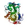
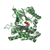
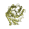
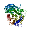
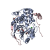
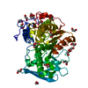
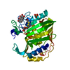
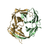
| Title | Crystal structure of recombinant glucuronoyl esterase from Sporotrichum thermophile determined at 1.55 A resolution | ||||||
 Components Components | 4-O-methyl-glucuronoyl methylesterase | ||||||
 Keywords Keywords | HYDROLASE / Alpha/Beta Hydrolase / 3-layer alpha/beta/alpha sandwich / Rossmann Fold / Glucuronoyl esterase | ||||||
| Function / homology |  Function and homology information Function and homology information(4-O-methyl)-D-glucuronate-lignin esterase / lignin catabolic process / carboxylic ester hydrolase activity / extracellular region Similarity search - Function | ||||||
| Biological species |  Myceliophthora thermophila (fungus) Myceliophthora thermophila (fungus) | ||||||
| Method |  X-RAY DIFFRACTION / SYNCHROTRON / MOLECULAR REPLACEMENT / Resolution: 1.55 Å X-RAY DIFFRACTION / SYNCHROTRON / MOLECULAR REPLACEMENT / Resolution: 1.55 Å | ||||||
 Authors Authors | Charavgi, M.D. / Dimarogona, M. / Topakas, E. / Christakopoulos, P. / Chrysina, E.D. | ||||||
 Citation Citation | Journal: Acta Crystallogr.,Sect.D / Year: 2013 Title: The structure of a novel glucuronoyl esterase from Myceliophthora thermophila gives new insights into its role as a potential biocatalyst. Authors: Charavgi, M.D. / Dimarogona, M. / Topakas, E. / Christakopoulos, P. / Chrysina, E.D. | ||||||
| History |
|
- Structure visualization
| Structure viewer | Molecule: MolmilJmol/JSmol |
|---|
- Downloads & links
-Download
| PDBx/mmCIF format | 4g4g.cif.gz | 169.4 KB | Display | PDBx/mmCIF format |
|---|---|---|---|---|
| PDB format | pdb4g4g.ent.gz | 131.7 KB | Display | PDB format |
| PDBx/mmJSON format | 4g4g.json.gz | Tree view | PDBx/mmJSON format | |
| Others |  Other downloads Other downloads |
-Validation report
| Arichive directory | https://data.pdbj.org/pub/pdb/validation_reports/g4/4g4gftp://data.pdbj.org/pub/pdb/validation_reports/g4/4g4g | HTTPS FTP |
|---|
-Related structure data
| Related structure data |  4g4iC  4g4jC  3picS C: citing same article ( S: Starting model for refinement |
|---|---|
| Similar structure data |


-Links
 PDBj
PDBj- Assembly
| Deposited unit | 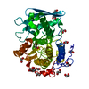
| ||||||||
|---|---|---|---|---|---|---|---|---|---|
| 1 |
| ||||||||
| Unit cell |
|
-Components
| #1: Protein | Mass: 46062.168 Da / Num. of mol.: 1 Source method: isolated from a genetically manipulated source Source: (gene. exp.) Myceliophthora thermophila (fungus) / Strain: ATCC 42464 / Gene: MYCTH_55568 / Plasmid: pPICZalphaC / Production host: Komagataella pastoris (fungus) / Strain (production host): X-33References: UniProt: G2QJR6, Hydrolases; Acting on ester bonds; Carboxylic-ester hydrolases | ||||||||
|---|---|---|---|---|---|---|---|---|---|
| #2: Chemical | ChemComp-EDO /   Mass: 62.068 Da / Num. of mol.: 14 / Source method: obtained synthetically / Formula: C2H6O2 Mass: 62.068 Da / Num. of mol.: 14 / Source method: obtained synthetically / Formula: C2H6O2#3: Chemical |   Mass: 92.094 Da / Num. of mol.: 2 / Source method: obtained synthetically / Formula: C3H8O3 Mass: 92.094 Da / Num. of mol.: 2 / Source method: obtained synthetically / Formula: C3H8O3#4: Water | ChemComp-HOH / |  Mass: 18.015 Da / Num. of mol.: 412 / Source method: isolated from a natural source / Formula: H2O Mass: 18.015 Da / Num. of mol.: 412 / Source method: isolated from a natural source / Formula: H2OHas protein modification | Y | Sequence details | THIS SEGMENT CORRESPOND | |
-Experimental details
-Experiment
| Experiment | Method: X-RAY DIFFRACTION / Number of used crystals: 1 |
|---|
- Sample preparation
| Crystal | Density Matthews: 2.2 Å3/Da / Density % sol: 44.08 % |
|---|---|
| Crystal grow | Temperature: 289 K / Method: vapor diffusion, sitting drop / pH: 8 Details: 30% PEG 3350, 0.1M Tris(hydroxymethyl)aminomethane hydrochloride, pH 8.0, VAPOR DIFFUSION, SITTING DROP, temperature 289.0K |
-Data collection
| Diffraction | Mean temperature: 100 K |
|---|---|
| Diffraction source | Source: SYNCHROTRON / Site: EMBL/DESY, HAMBURG  / Beamline: X13 / Wavelength: 0.8123 Å / Beamline: X13 / Wavelength: 0.8123 Å |
| Detector | Type: MAR CCD 165 mm / Detector: CCD / Date: Nov 23, 2010 / Details: mirrors |
| Radiation | Monochromator: Si (111) horizontally focussing / Protocol: SINGLE WAVELENGTH / Monochromatic (M) / Laue (L): M / Scattering type: x-ray |
| Radiation wavelength | Wavelength: 0.8123 Å / Relative weight: 1 |
| Reflection | Resolution: 1.55→19.45 Å / Num. all: 51271 / Num. obs: 51271 / % possible obs: 94.5 % / Observed criterion σ(F): 0 / Observed criterion σ(I): 0 / Redundancy: 3.7 % / Biso Wilson estimate: 13.5 Å2 / Rmerge(I) obs: 0.069 / Net I/σ(I): 9.5 |
| Reflection shell | Resolution: 1.55→1.63 Å / Redundancy: 3.5 % / Rmerge(I) obs: 0.498 / Mean I/σ(I) obs: 2.5 / Num. unique all: 7498 / % possible all: 95.9 |
- Processing
| Software |
| ||||||||||||||||||||||||||||||||||||||||||||||||||||||||||||
|---|---|---|---|---|---|---|---|---|---|---|---|---|---|---|---|---|---|---|---|---|---|---|---|---|---|---|---|---|---|---|---|---|---|---|---|---|---|---|---|---|---|---|---|---|---|---|---|---|---|---|---|---|---|---|---|---|---|---|---|---|---|
| Refinement | Method to determine structure: MOLECULAR REPLACEMENT Starting model: PDB ENTRY 3PIC Resolution: 1.55→19.36 Å / Cor.coef. Fo:Fc: 0.945 / Cor.coef. Fo:Fc free: 0.917 / SU B: 4.308 / SU ML: 0.071 / Cross valid method: THROUGHOUT / σ(F): 0 / ESU R: 0.134 / ESU R Free: 0.105 / Stereochemistry target values: MAXIMUM LIKELIHOOD / Details: HYDROGENS HAVE BEEN USED IF PRESENT IN THE INPUT
| ||||||||||||||||||||||||||||||||||||||||||||||||||||||||||||
| Solvent computation | Ion probe radii: 0.8 Å / Shrinkage radii: 0.8 Å / VDW probe radii: 1.2 Å / Solvent model: MASK | ||||||||||||||||||||||||||||||||||||||||||||||||||||||||||||
| Displacement parameters | Biso mean: 17.129 Å2
| ||||||||||||||||||||||||||||||||||||||||||||||||||||||||||||
| Refine analyze | Luzzati coordinate error obs: 0.2138 Å | ||||||||||||||||||||||||||||||||||||||||||||||||||||||||||||
| Refinement step | Cycle: LAST / Resolution: 1.55→19.36 Å
| ||||||||||||||||||||||||||||||||||||||||||||||||||||||||||||
| Refine LS restraints |
| ||||||||||||||||||||||||||||||||||||||||||||||||||||||||||||
| LS refinement shell | Resolution: 1.55→1.59 Å / Total num. of bins used: 20
|
