 Movie
Movie Controller


+ Open data

- Basic information
| Entry | Database: PDB / ID: 1b9m | ||||||
|---|---|---|---|---|---|---|---|
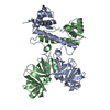
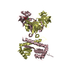
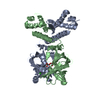
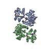
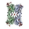
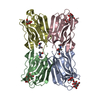
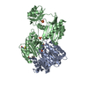
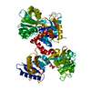
| Title | REGULATOR FROM ESCHERICHIA COLI | ||||||
 Components Components | PROTEIN (MODE) | ||||||
 Keywords Keywords | TRANSCRIPTION / DNA-BINDING / GENE REGULATION / WINGED HELIX TURN HELIX / MOLYBDATE / OB FOLD | ||||||
| Function / homology |  Function and homology information Function and homology informationModE complex / negative regulation of DNA-templated transcription initiation / molybdate ion transport / molybdenum ion binding / cis-regulatory region sequence-specific DNA binding / negative regulation of DNA-templated transcription / regulation of DNA-templated transcription / positive regulation of DNA-templated transcription / cytosol Similarity search - Function | ||||||
| Biological species |  | ||||||
| Method |  X-RAY DIFFRACTION / SYNCHROTRON / MAD / Resolution: 1.75 Å X-RAY DIFFRACTION / SYNCHROTRON / MAD / Resolution: 1.75 Å | ||||||
 Authors Authors | Hall, D.R. / Gourley, D.G. / Hunter, W.N. | ||||||
 Citation Citation | Journal: EMBO J. / Year: 1999 Title: The high-resolution crystal structure of the molybdate-dependent transcriptional regulator (ModE) from Escherichia coli: a novel combination of domain folds. Authors: Hall, D.R. / Gourley, D.G. / Leonard, G.A. / Duke, E.M. / Anderson, L.A. / Boxer, D.H. / Hunter, W.N. #1: Journal: Acta Crystallogr.,Sect.D / Year: 1999 Title: Two crystal forms of ModE, the molybdate-dependent transcriptional regulator from Escherichia coli. Authors: Hall, D.R. / Gourley, D.G. / Duke, E.M. / Leonard, G.A. / Anderson, L.A. / Pau, R.N. / Boxer, D.H. / Hunter, W.N. #2: Journal: Eur.J.Biochem. / Year: 1997 Title: Characterisation of the molybdenum-responsive ModE regulatory protein and its binding to the promoter region of the modABCD (molybdenum transport) operon of Escherichia coli. Authors: Anderson, L.A. / Palmer, T. / Price, N.C. / Bornemann, S. / Boxer, D.H. / Pau, R.N. | ||||||
| History |
|
- Structure visualization
| Structure viewer | Molecule: MolmilJmol/JSmol |
|---|
- Downloads & links
-Download
| PDBx/mmCIF format | 1b9m.cif.gz | 119.2 KB | Display | PDBx/mmCIF format |
|---|---|---|---|---|
| PDB format | pdb1b9m.ent.gz | 91.3 KB | Display | PDB format |
| PDBx/mmJSON format | 1b9m.json.gz | Tree view | PDBx/mmJSON format | |
| Others |  Other downloads Other downloads |
-Validation report
| Arichive directory | https://data.pdbj.org/pub/pdb/validation_reports/b9/1b9mftp://data.pdbj.org/pub/pdb/validation_reports/b9/1b9m | HTTPS FTP |
|---|
-Related structure data


-Links
 PDBj
PDBj- Assembly
| Deposited unit | 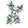
| ||||||||
|---|---|---|---|---|---|---|---|---|---|
| 1 |
| ||||||||
| Unit cell |
|
-Components
| #1: Protein | Mass: 28731.100 Da / Num. of mol.: 2 Source method: isolated from a genetically manipulated source Source: (gene. exp.) #2: Chemical | ChemComp-NI / |   Mass: 58.693 Da / Num. of mol.: 1 / Source method: obtained synthetically / Formula: Ni Mass: 58.693 Da / Num. of mol.: 1 / Source method: obtained synthetically / Formula: Ni#3: Water | ChemComp-HOH / |  Mass: 18.015 Da / Num. of mol.: 426 / Source method: isolated from a natural source / Formula: H2O Mass: 18.015 Da / Num. of mol.: 426 / Source method: isolated from a natural source / Formula: H2OHas protein modification | Y | |
|---|
-Experimental details
-Experiment
| Experiment | Method: X-RAY DIFFRACTION / Number of used crystals: 2 |
|---|
- Sample preparation
| Crystal | Density Matthews: 3 Å3/Da / Density % sol: 59 % Description: THESE ARE THE PARAMETERS FOR THE DATA SET USED FOR REFINEMENT. THE DATA USED FOR STRUCTURE SOLUTION ARE ALSO SUPPLIED. | ||||||||||||||||||||||||||||||
|---|---|---|---|---|---|---|---|---|---|---|---|---|---|---|---|---|---|---|---|---|---|---|---|---|---|---|---|---|---|---|---|
| Crystal grow | pH: 6.5 / Details: pH 6.5 | ||||||||||||||||||||||||||||||
| Crystal grow | *PLUS Temperature: 277 K / pH: 60 / Method: vapor diffusion, hanging drop | ||||||||||||||||||||||||||||||
| Components of the solutions | *PLUS
|
-Data collection
| Diffraction | Mean temperature: 100 K | |||||||||
|---|---|---|---|---|---|---|---|---|---|---|
| Diffraction source | Source: SYNCHROTRON / Site: ESRF  / Beamline: BM14 / Wavelength: 0.8855, 0.9310 / Beamline: BM14 / Wavelength: 0.8855, 0.9310 | |||||||||
| Detector | Type: PRINCETON 2K / Detector: CCD / Date: Apr 1, 1998 | |||||||||
| Radiation | Protocol: MAD / Monochromatic (M) / Laue (L): M / Scattering type: x-ray | |||||||||
| Radiation wavelength |
| |||||||||
| Reflection | Resolution: 1.75→24.15 Å / Num. obs: 205040 / % possible obs: 80.2 % / Redundancy: 3 % / Biso Wilson estimate: 31.43 Å2 / Rmerge(I) obs: 0.055 / Net I/σ(I): 31.8 | |||||||||
| Reflection shell | Resolution: 1.75→1.78 Å / Redundancy: 1 % / Rmerge(I) obs: 0.368 / Mean I/σ(I) obs: 2.9 / % possible all: 66.4 | |||||||||
| Reflection | *PLUS Num. obs: 53298 / Num. measured all: 205040 | |||||||||
| Reflection shell | *PLUS % possible obs: 66.4 % |
- Processing
| Software |
| ||||||||||||||||||||||||||||||||||||||||||||||||||||||||||||||||||||||||||||||||||||
|---|---|---|---|---|---|---|---|---|---|---|---|---|---|---|---|---|---|---|---|---|---|---|---|---|---|---|---|---|---|---|---|---|---|---|---|---|---|---|---|---|---|---|---|---|---|---|---|---|---|---|---|---|---|---|---|---|---|---|---|---|---|---|---|---|---|---|---|---|---|---|---|---|---|---|---|---|---|---|---|---|---|---|---|---|---|
| Refinement | Method to determine structure: MAD / Resolution: 1.75→24.15 Å / SU B: 2.94753 / SU ML: 0.09435 / Cross valid method: THROUGHOUT / σ(F): 0 / ESU R: 0.16091 / ESU R Free: 0.15609 Details: SULPHUR ATOMS OF ALL METHIONINE RESIDUES WERE REPLACED BY SELENIUM ATOMS.
| ||||||||||||||||||||||||||||||||||||||||||||||||||||||||||||||||||||||||||||||||||||
| Displacement parameters | Biso mean: 41.8 Å2 | ||||||||||||||||||||||||||||||||||||||||||||||||||||||||||||||||||||||||||||||||||||
| Refinement step | Cycle: LAST / Resolution: 1.75→24.15 Å
| ||||||||||||||||||||||||||||||||||||||||||||||||||||||||||||||||||||||||||||||||||||
| Refine LS restraints |
|
