 Movie
Movie Controller


+ Open data

- Basic information
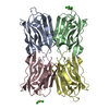
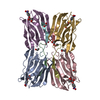
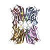
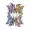
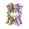
| Entry | Database: PDB / ID: 1vbo | |||||||||
|---|---|---|---|---|---|---|---|---|---|---|
| Title | Crystal structure of artocarpin-mannotriose complex | |||||||||
 Components Components | artocarpin | |||||||||
 Keywords Keywords | PLANT PROTEIN / beta-prism / mannose-specific / lectin / jacalin-like | |||||||||
| Function / homology |  Function and homology information Function and homology information | |||||||||
| Biological species |   Artocarpus integer (chempedak) Artocarpus integer (chempedak) | |||||||||
| Method |  X-RAY DIFFRACTION / MOLECULAR REPLACEMENT / Resolution: 2.35 Å X-RAY DIFFRACTION / MOLECULAR REPLACEMENT / Resolution: 2.35 Å | |||||||||
 Authors Authors | Jeyaprakash, A.A. / Srivastav, A. / Surolia, A. / Vijayan, M. | |||||||||
 Citation Citation | Journal: J.Mol.Biol. / Year: 2004 Title: Structural basis for the carbohydrate specificities of artocarpin: variation in the length of a loop as a strategy for generating ligand specificity Authors: Jeyaprakash, A.A. / Srivastav, A. / Surolia, A. / Vijayan, M. #1: Journal: J.Mol.Biol. / Year: 2002Title: Crystal structures of artocarpin, a Moraceae lectin with mannose specificity, and its complex with methyl-alpha-D-mannose: implications to the generation of carbohydrate specificity Authors: Pratap, J.V. / Jeyaprakash, A.A. / Rani, P.G. / Sekar, K. / Surolia, A. / Vijayan, M. #2: Journal: NAT.STRUCT.BIOL. / Year: 1996Title: A novel mode of carbohydrate recognition in jacalin, a Moraceae plant lectin with a beta-prism fold Authors: Sankaranarayanan, R. / Sekar, K. / Banerjee, R. / Sharma, V. / Surolia, A. / Vijayan, M. #3: Journal: J.Mol.Biol. / Year: 2003Title: Structural basis of the carbohydrate specificities of jacalin: an X-ray and modeling study Authors: Jeyaprakash, A.A. / Katiyar, S. / Swaminathan, C.P. / Sekar, K. / Surolia, A. / Vijayan, M. | |||||||||
| History |
|
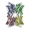
- Structure visualization
| Structure viewer | Molecule: MolmilJmol/JSmol |
|---|
- Downloads & links
-Download
| PDBx/mmCIF format | 1vbo.cif.gz | 249.8 KB | Display | PDBx/mmCIF format |
|---|---|---|---|---|
| PDB format | pdb1vbo.ent.gz | 203.3 KB | Display | PDB format |
| PDBx/mmJSON format | 1vbo.json.gz | Tree view | PDBx/mmJSON format | |
| Others |  Other downloads Other downloads |
-Validation report
| Arichive directory | https://data.pdbj.org/pub/pdb/validation_reports/vb/1vboftp://data.pdbj.org/pub/pdb/validation_reports/vb/1vbo | HTTPS FTP |
|---|
-Related structure data
| Related structure data | 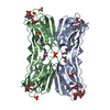 1vbpC 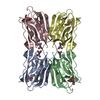 1j4uS S: Starting model for refinement C: citing same article ( |
|---|---|
| Similar structure data |


-Links
 PDBj
PDBj


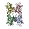
- Assembly
| Deposited unit | 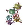
| ||||||||
|---|---|---|---|---|---|---|---|---|---|
| 1 | 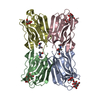
| ||||||||
| 2 | 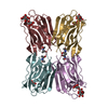
| ||||||||
| Unit cell |
|
-Components
| #1: Protein | Mass: 16112.988 Da / Num. of mol.: 8 / Source method: isolated from a natural source / Source: (natural) Artocarpus integer (chempedak) / Tissue: seeds / References: UniProt: Q7M1T4#2: Polysaccharide | alpha-D-mannopyranose-(1-3)-[alpha-D-mannopyranose-(1-6)]alpha-D-mannopyranose Source method: isolated from a genetically manipulated source #3: Sugar |   Type: D-saccharide, alpha linking / Mass: 180.156 Da / Num. of mol.: 2 Type: D-saccharide, alpha linking / Mass: 180.156 Da / Num. of mol.: 2Source method: isolated from a genetically manipulated source Formula: C6H12O6 #4: Water | ChemComp-HOH / |  Mass: 18.015 Da / Num. of mol.: 490 / Source method: isolated from a natural source / Formula: H2O Mass: 18.015 Da / Num. of mol.: 490 / Source method: isolated from a natural source / Formula: H2O |
|---|
-Experimental details
-Experiment
| Experiment | Method: X-RAY DIFFRACTION / Number of used crystals: 1 |
|---|
- Sample preparation
| Crystal | Density Matthews: 2.15 Å3/Da / Density % sol: 42.22 % |
|---|---|
| Crystal grow | Temperature: 300 K / Method: vapor diffusion, hanging drop / pH: 7.4 Details: PEG4000, PEG1450, Phosphate buffer, pH 7.4, VAPOR DIFFUSION, HANGING DROP, temperature 300K |
-Data collection
| Diffraction | Mean temperature: 278 K |
|---|---|
| Diffraction source | Source: ROTATING ANODE / Type: RIGAKU RU200 / Wavelength: 1.5418 Å |
| Detector | Type: MARRESEARCH / Detector: IMAGE PLATE / Date: Jun 10, 2003 / Details: OSMIC mirrors |
| Radiation | Monochromator: OSMIC mirrors / Protocol: SINGLE WAVELENGTH / Monochromatic (M) / Laue (L): M / Scattering type: x-ray |
| Radiation wavelength | Wavelength: 1.5418 Å / Relative weight: 1 |
| Reflection | Resolution: 2.35→20 Å / Num. all: 48504 / Num. obs: 48504 / % possible obs: 99.2 % / Observed criterion σ(F): 0 / Observed criterion σ(I): 0 / Redundancy: 2.8 % / Biso Wilson estimate: 31.9 Å2 / Rmerge(I) obs: 0.104 / Net I/σ(I): 8.9 |
| Reflection shell | Resolution: 2.35→2.43 Å / Rmerge(I) obs: 0.498 / Num. unique all: 4824 / % possible all: 99.3 |
- Processing
| Software |
| ||||||||||||||||||||||||||||||||||||
|---|---|---|---|---|---|---|---|---|---|---|---|---|---|---|---|---|---|---|---|---|---|---|---|---|---|---|---|---|---|---|---|---|---|---|---|---|---|
| Refinement | Method to determine structure: MOLECULAR REPLACEMENT Starting model: 1J4U Resolution: 2.35→19.85 Å / Rfactor Rfree error: 0.005 / Data cutoff high absF: 1485698.72 / Data cutoff low absF: 0 / Isotropic thermal model: RESTRAINED / Cross valid method: THROUGHOUT / σ(F): 0 / Stereochemistry target values: Engh & Huber
| ||||||||||||||||||||||||||||||||||||
| Solvent computation | Solvent model: FLAT MODEL / Bsol: 32.311 Å2 / ksol: 0.298422 e/Å3 | ||||||||||||||||||||||||||||||||||||
| Displacement parameters | Biso mean: 39.9 Å2
| ||||||||||||||||||||||||||||||||||||
| Refine analyze |
| ||||||||||||||||||||||||||||||||||||
| Refinement step | Cycle: LAST / Resolution: 2.35→19.85 Å
| ||||||||||||||||||||||||||||||||||||
| Refine LS restraints |
| ||||||||||||||||||||||||||||||||||||
| LS refinement shell | Resolution: 2.35→2.5 Å / Rfactor Rfree error: 0.016 / Total num. of bins used: 6
| ||||||||||||||||||||||||||||||||||||
| Xplor file |
|
