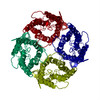
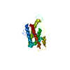
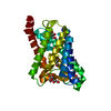
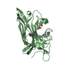
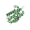
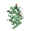
ジャーナル: FEBS Lett / 年: 2001 タイトル: A refined structure of human aquaporin-1. 著者: B L de Groot / A Engel / H Grubmüller / 要旨: A refined structure of the human water channel aquaporin-1 is presented. The model rests on the high resolution X-ray structure of the homologous bacterial glycerol transporter GlpF, electron ...A refined structure of the human water channel aquaporin-1 is presented. The model rests on the high resolution X-ray structure of the homologous bacterial glycerol transporter GlpF, electron crystallographic data at 3.8 A resolution and a multiple sequence alignment of the aquaporin superfamily. The crystallographic R and free R values (36.7% and 37.8%) for the refined structure are significantly lower than for previous models. Improved geometry and enhanced stability in molecular dynamics simulations demonstrate a significant improvement of the aquaporin-1 structure. Comparison with previous aquaporin-1 models shows significant differences, not only in the loop regions, but also in the core of the water channel.
#1: ジャーナル: J Mol Biol / 年: 2000 タイトル: The fold of human aquaporin 1. 著者: B L de Groot / J B Heymann / A Engel / K Mitsuoka / Y Fujiyoshi / H Grubmüller / 要旨: The fold of human aquaporin 1 is determined from cryo-electron microscopic data at 4.5 A resolution. The monomeric structure consists of two transmembrane triple helices arranged around a pseudo-2- ...The fold of human aquaporin 1 is determined from cryo-electron microscopic data at 4.5 A resolution. The monomeric structure consists of two transmembrane triple helices arranged around a pseudo-2-fold axis connected by a long flexible extracellular loop. Each triplet contains between its second and third helix a functional loop containing the highly conserved fingerprint NPA motif. These functional loops are assumed to fold inwards between the two triplets, thereby forming the heart of the water channel. The helix topology was determined from the directionality pattern of each of the six transmembrane helices with respect to the membrane, together with constraints defined by the sequence and atomic force microscopy data. The directionality of the helices was determined by collecting the best-fitting orientations resulting from a search through the three-dimensional experimental map for a large number of alpha-helical fragments. Tests on cryo-electron crystallographic bacteriorhodopsin data suggest that our method is generally applicable to determine the topology of helical proteins for which only medium-resolution electron microscopy data are available.
開始モデル: THE MODEL WAS DERIVED USING ELECTRON DIFFRACTION DATA ON 2D CRYSTALS 解像度: 3.54→30.04 Å / Rfactor Rfree error: 0.017 / Isotropic thermal model: GROUP / 交差検証法: THROUGHOUT / σ(F): 0
 ムービー
ムービー コントローラー


 データを開く
 基本情報
基本情報 要素
要素 キーワード
キーワード 機能・相同性情報
 HOMO SAPIENS (ヒト)
 データ登録者
 引用
引用
 構造の表示
構造の表示 ダウンロードとリンク
 その他のダウンロード
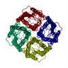

 PDBj
PDBj

 集合体
集合体
 試料調製
試料調製 FIELD EMISSION GUN / 加速電圧: 300 kV / 照射モード: FLOOD BEAM
 解析
解析