 Movie
Movie Controller


+ Open data

- Basic information
| Entry | Database: PDB / ID: 1ih5 | |||||||||
|---|---|---|---|---|---|---|---|---|---|---|
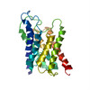
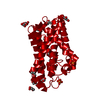
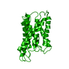
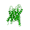
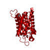
| Title | CRYSTAL STRUCTURE OF AQUAPORIN-1 | |||||||||
 Components Components | AQUAPORIN-1 | |||||||||
 Keywords Keywords | MEMBRANE PROTEIN / WATER CHANNEL / TWO-DIMENSIONAL CRYSTAL | |||||||||
| Function / homology |  Function and homology information Function and homology informationmetanephric descending thin limb development / metanephric proximal straight tubule development / metanephric proximal convoluted tubule segment 2 development / metanephric glomerulus vasculature development / nitric oxide transmembrane transporter activity / hydrogen peroxide channel activity / cerebrospinal fluid secretion / lipid digestion / renal water transport / corticotropin secretion ...metanephric descending thin limb development / metanephric proximal straight tubule development / metanephric proximal convoluted tubule segment 2 development / metanephric glomerulus vasculature development / nitric oxide transmembrane transporter activity / hydrogen peroxide channel activity / cerebrospinal fluid secretion / lipid digestion / renal water transport / corticotropin secretion / cellular response to salt stress / carbon dioxide transmembrane transport / secretory granule organization / carbon dioxide transmembrane transporter activity / glycerol transmembrane transporter activity / cellular response to mercury ion / water transmembrane transporter activity / positive regulation of saliva secretion / Passive transport by Aquaporins / pancreatic juice secretion / lateral ventricle development / establishment or maintenance of actin cytoskeleton polarity / glycerol transmembrane transport / intracellular water homeostasis / intracellularly cGMP-activated cation channel activity / renal water absorption / potassium ion transmembrane transporter activity / transepithelial water transport / water channel activity / ankyrin-1 complex / ammonium transmembrane transport / ammonium channel activity / fibroblast migration / glomerular filtration / camera-type eye morphogenesis / multicellular organismal-level water homeostasis / water transport / hyperosmotic response / cellular homeostasis / cellular hyperosmotic response / odontogenesis / cell volume homeostasis / positive regulation of fibroblast migration / nitric oxide transport / brush border / cellular response to dexamethasone stimulus / potassium channel activity / establishment of localization in cell / renal water homeostasis / ephrin receptor binding / transmembrane transporter activity / cellular response to retinoic acid / cellular response to nitric oxide / sensory perception of pain / basal plasma membrane / cellular response to cAMP / cellular response to copper ion / potassium ion transport / Developmental Lineage of Pancreatic Ductal Cells / carbon dioxide transport / wound healing / brush border membrane / Erythrocytes take up oxygen and release carbon dioxide / Erythrocytes take up carbon dioxide and release oxygen / cellular response to mechanical stimulus / sarcolemma / positive regulation of fibroblast proliferation / cellular response to hydrogen peroxide / positive regulation of angiogenesis / apical part of cell / cellular response to UV / Vasopressin regulates renal water homeostasis via Aquaporins / nuclear membrane / cellular response to hypoxia / defense response to Gram-negative bacterium / basolateral plasma membrane / apical plasma membrane / axon / negative regulation of apoptotic process / extracellular exosome / identical protein binding / nucleus / plasma membrane / cytoplasm Similarity search - Function | |||||||||
| Biological species |  Homo sapiens (human) Homo sapiens (human) | |||||||||
| Method | ELECTRON CRYSTALLOGRAPHY / electron crystallography / cryo EM / Resolution: 3.7 Å | |||||||||
 Authors Authors | Ren, G. / Reddy, V.S. / Cheng, A. / Melnyk, P. / Mitra, A.K. | |||||||||
 Citation Citation | Journal: Proc Natl Acad Sci U S A / Year: 2001 Title: Visualization of a water-selective pore by electron crystallography in vitreous ice. Authors: G Ren / V S Reddy / A Cheng / P Melnyk / A K Mitra /  Abstract: The water-selective pathway through the aquaporin-1 membrane channel has been visualized by fitting an atomic model to a 3.7-A resolution three-dimensional density map. This map was determined by ...The water-selective pathway through the aquaporin-1 membrane channel has been visualized by fitting an atomic model to a 3.7-A resolution three-dimensional density map. This map was determined by analyzing images and electron diffraction patterns of lipid-reconstituted two-dimensional crystals of aquaporin-1 preserved in vitrified buffer in the absence of any additive. The aqueous pathway is characterized by a size-selective pore that is approximately 4.0 +/- 0.5A in diameter, spans a length of approximately 18A, and bends by approximately 25 degrees as it traverses the bilayer. This narrow pore is connected by wide, funnel-shaped openings at the extracellular and cytoplasmic faces. The size-selective pore is outlined mostly by hydrophobic residues, resulting in a relatively inert pathway conducive to diffusion-limited water flow. The apex of the curved pore is close to the locations of the in-plane pseudo-2-fold symmetry axis that relates the N- and C-terminal halves and the conserved, functionally important N76 and N192 residues. #1: Journal: J.Mol.Biol. / Year: 2000Title: Three-Dimensional Fold of the Human Aqp1 Water Channel Determined at 4 A Resolution by Electron Crystallography of Two-Dimensional Crystals Embedded in Ice Authors: Ren, G. / Cheng, A. / Reddy, V. / Melnyk, P. / Mitra, A.K. #2: Journal: J.Struct.Biol. / Year: 2000Title: Polymorphism in the Packing of Aquaporin-1 Tetramers in 2-D Crystals Authors: Ren, G. / Cheng, A. / Melnyk, P. / Mitra, A.K. #3: Journal: Nature / Year: 1997Title: Three-Dimensional Organization of a Human Water Channel Authors: Cheng, A. / Van Hoek, A.N. / Yeager, M. / Verkman, A.S. / Mitra, A.K. | |||||||||
| History |
|
- Structure visualization
| Movie |
Movie viewer |
|---|---|
| Structure viewer | Molecule: MolmilJmol/JSmol |
- Downloads & links
-Download
| PDBx/mmCIF format | 1ih5.cif.gz | 48.3 KB | Display | PDBx/mmCIF format |
|---|---|---|---|---|
| PDB format | pdb1ih5.ent.gz | 34.5 KB | Display | PDB format |
| PDBx/mmJSON format | 1ih5.json.gz | Tree view | PDBx/mmJSON format | |
| Others |  Other downloads Other downloads |
-Validation report
| Arichive directory | https://data.pdbj.org/pub/pdb/validation_reports/ih/1ih5ftp://data.pdbj.org/pub/pdb/validation_reports/ih/1ih5 | HTTPS FTP |
|---|
-Related structure data
| Related structure data | |
|---|---|
| Similar structure data |
-Links
 PDBj
PDBj

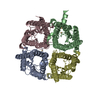
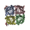
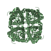
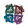
- Assembly
| Deposited unit | 
| ||||||||
|---|---|---|---|---|---|---|---|---|---|
| 1 | 
| ||||||||
| Unit cell |
|
-Components
| #1: Protein | Mass: 28549.914 Da / Num. of mol.: 1 / Source method: isolated from a natural source / Source: (natural) Homo sapiens (human) / Cell: RED-BLOOD CELL / Cellular location: MEMBRANE / References: UniProt: P29972 |
|---|
-Experimental details
-Experiment
| Experiment | Method: ELECTRON CRYSTALLOGRAPHY |
|---|---|
| EM experiment | Aggregation state: 2D ARRAY / 3D reconstruction method: electron crystallography |
- Sample preparation
| Component | Name: aquaporin / Type: COMPLEX | ||||||||||||||||||||||||||||||||||||||||||||||||
|---|---|---|---|---|---|---|---|---|---|---|---|---|---|---|---|---|---|---|---|---|---|---|---|---|---|---|---|---|---|---|---|---|---|---|---|---|---|---|---|---|---|---|---|---|---|---|---|---|---|
| Specimen | Embedding applied: NO / Shadowing applied: NO / Staining applied: NO / Vitrification applied: YES | ||||||||||||||||||||||||||||||||||||||||||||||||
| Crystal grow | *PLUS pH: 7.2 / Method: unknown | ||||||||||||||||||||||||||||||||||||||||||||||||
| Components of the solutions | *PLUS
|
-Data collection
| Microscopy | Model: FEI/PHILIPS CM200FEG |
|---|---|
| Electron gun | Electron source:  FIELD EMISSION GUN / Illumination mode: FLOOD BEAM FIELD EMISSION GUN / Illumination mode: FLOOD BEAM |
| Electron lens | Mode: DIFFRACTION |
| Detector | Date: Jan 1, 1999 |
| Radiation | Protocol: SINGLE WAVELENGTH / Monochromatic (M) / Laue (L): M / Scattering type: electron |
| Radiation wavelength | Relative weight: 1 |
| Reflection | Biso Wilson estimate: 38.1 Å2 |
| Reflection | *PLUS Highest resolution: 3.7 Å / Num. obs: 19839 / % possible obs: 63 % / Num. measured all: 48037 |
- Processing
| Software | Name: X-PLOR / Classification: refinement | ||||||||||||||||||||
|---|---|---|---|---|---|---|---|---|---|---|---|---|---|---|---|---|---|---|---|---|---|
| 3D reconstruction | Resolution: 3.7 Å / Resolution method: DIFFRACTION PATTERN/LAYERLINES | ||||||||||||||||||||
| Refinement | Resolution: 3.7→24 Å / Isotropic thermal model: ISOTROPIC / Cross valid method: THROUGHOUT / σ(F): 2 Details: POSITIONAL REFINEMENT USING X-PLOR. REFINEMENT MONITORED BY RFREE AND PHIFREE (<| CALCLD. PHASE - EXPTL. OBSERVED PHASE|>). PSEUDO 2-FOLD SYMMETRY IN THE PLANE OF THE BILAYER RELATES THE N- AND C-TERMINAL HALVES
| ||||||||||||||||||||
| Displacement parameters | Biso mean: 47.8 Å2 | ||||||||||||||||||||
| Refinement step | Cycle: LAST / Resolution: 3.7→24 Å
|
