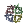
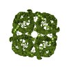
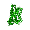
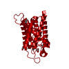
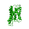
Journal: Nature / Year: 2000 Title: Structural determinants of water permeation through aquaporin-1. Authors: K Murata / K Mitsuoka / T Hirai / T Walz / P Agre / J B Heymann / A Engel / Y Fujiyoshi / Abstract: Human red cell AQP1 is the first functionally defined member of the aquaporin family of membrane water channels. Here we describe an atomic model of AQP1 at 3.8A resolution from electron ...Human red cell AQP1 is the first functionally defined member of the aquaporin family of membrane water channels. Here we describe an atomic model of AQP1 at 3.8A resolution from electron crystallographic data. Multiple highly conserved amino-acid residues stabilize the novel fold of AQP1. The aqueous pathway is lined with conserved hydrophobic residues that permit rapid water transport, whereas the water selectivity is due to a constriction of the pore diameter to about 3 A over a span of one residue. The atomic model provides a possible molecular explanation to a longstanding puzzle in physiology-how membranes can be freely permeable to water but impermeable to protons.
History
Deposition
Sep 7, 2000
Deposition site: RCSB / Processing site: RCSB
Revision 1.0
Oct 18, 2000
Provider: repository / Type: Initial release
Revision 1.1
Apr 27, 2008
Group: Version format compliance
Revision 1.2
Jul 13, 2011
Group: Derived calculations / Version format compliance
Resolution: 3.8 Å / Resolution method: DIFFRACTION PATTERN/LAYERLINES
Refinement
Resolution: 3.8→6 Å / σ(F): 1 / σ(I): 0 / Stereochemistry target values: Engh & Huber Details: For refinement with phase restraint, we used the structure factors merged from both electron diffraction patterns and images. Thus in this file we included the structure factors with phases ...Details: For refinement with phase restraint, we used the structure factors merged from both electron diffraction patterns and images. Thus in this file we included the structure factors with phases used in the refinement. The observed phases were labelled as calculated phases here.
Rfactor
Num. reflection
% reflection
Selection details
Rfree
0.417
124
0.036 %
RANDOM
Rwork
0.399
-
-
-
all
0.406
1846
-
-
obs
0.406
1846
50.8 %
-
Refine analyze
Luzzati coordinate error obs: 0.85 Å
Refinement step
Cycle: LAST / Resolution: 3.8→6 Å
Protein
Nucleic acid
Ligand
Solvent
Total
Num. atoms
1661
0
0
0
1661
Refine LS restraints
Refine-ID
Type
Dev ideal
ELECTRONCRYSTALLOGRAPHY
x_bond_d
0.017
ELECTRONCRYSTALLOGRAPHY
x_angle_deg
2.794
LS refinement shell
Resolution: 3.8→3.93 Å / Total num. of bins used: 8
Rfactor
% reflection
Rfree
0.31
1.58 %
Rwork
0.424
-
obs
-
36.3 %
+
About Yorodumi
-
News
-
Feb 9, 2022. New format data for meta-information of EMDB entries
New format data for meta-information of EMDB entries
Version 3 of the EMDB header file is now the official format.
The previous official version 1.9 will be removed from the archive.
In the structure databanks used in Yorodumi, some data are registered as the other names, "COVID-19 virus" and "2019-nCoV". Here are the details of the virus and the list of structure data.
Jan 31, 2019. EMDB accession codes are about to change! (news from PDBe EMDB page)
EMDB accession codes are about to change! (news from PDBe EMDB page)
The allocation of 4 digits for EMDB accession codes will soon come to an end. Whilst these codes will remain in use, new EMDB accession codes will include an additional digit and will expand incrementally as the available range of codes is exhausted. The current 4-digit format prefixed with “EMD-” (i.e. EMD-XXXX) will advance to a 5-digit format (i.e. EMD-XXXXX), and so on. It is currently estimated that the 4-digit codes will be depleted around Spring 2019, at which point the 5-digit format will come into force.
The EM Navigator/Yorodumi systems omit the EMD- prefix.
Related info.:Q: What is EMD? / ID/Accession-code notation in Yorodumi/EM Navigator
Yorodumi is a browser for structure data from EMDB, PDB, SASBDB, etc.
This page is also the successor to EM Navigator detail page, and also detail information page/front-end page for Omokage search.
The word "yorodu" (or yorozu) is an old Japanese word meaning "ten thousand". "mi" (miru) is to see.
Related info.:EMDB / PDB / SASBDB / Comparison of 3 databanks / Yorodumi Search / Aug 31, 2016. New EM Navigator & Yorodumi / Yorodumi Papers / Jmol/JSmol / Function and homology information / Changes in new EM Navigator and Yorodumi
 Movie
Movie Controller

 Yorodumi
 Open data
 Basic information
 Components
 Keywords
 Function and homology information
 Homo sapiens (human)
 Authors
 Citation
 Structure visualization
 Downloads & links
 Other downloads
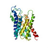


 PDBj
PDBj

 Assembly
 Sample preparation
 FIELD EMISSION GUN / Accelerating voltage: 300 kV / Illumination mode: FLOOD BEAM
 Processing