 Movie
Movie Controller

[English] 日本語
 Yorodumi
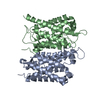
- PDB-3cn5: Crystal structure of the Spinach Aquaporin SoPIP2;1 S115E, S274E ... -
+ Open data

- Basic information
| Entry | Database: PDB / ID: 3cn5 | ||||||
|---|---|---|---|---|---|---|---|
| Title | Crystal structure of the Spinach Aquaporin SoPIP2;1 S115E, S274E mutant | ||||||
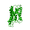
 Components Components | Aquaporin | ||||||
 Keywords Keywords | TRANSPORT PROTEIN / membrane protein / aquaporin / Transmembrane / Transport | ||||||
| Function / homology |  Function and homology information Function and homology information | ||||||
| Biological species |  Spinacia oleracea (spinach) Spinacia oleracea (spinach) | ||||||
| Method |  X-RAY DIFFRACTION / SYNCHROTRON / MOLECULAR REPLACEMENT / Resolution: 2.05 Å X-RAY DIFFRACTION / SYNCHROTRON / MOLECULAR REPLACEMENT / Resolution: 2.05 Å | ||||||
 Authors Authors | Nyblom, M. / Alfredsson, A. / Hallgren, K. / Hedfalk, K. / Neutze, R. / Tornroth-Horsefield, S. | ||||||
 Citation Citation | Journal: J.Mol.Biol. / Year: 2009 Title: Structural and functional analysis of SoPIP2;1 mutants adds insight into plant aquaporin gating. Authors: Nyblom, M. / Frick, A. / Wang, Y. / Ekvall, M. / Hallgren, K. / Hedfalk, K. / Neutze, R. / Tajkhorshid, E. / Tornroth-Horsefield, S. | ||||||
| History |
|
- Structure visualization
| Structure viewer | Molecule: MolmilJmol/JSmol |
|---|
- Downloads & links
-Download
| PDBx/mmCIF format | 3cn5.cif.gz | 60.7 KB | Display | PDBx/mmCIF format |
|---|---|---|---|---|
| PDB format | pdb3cn5.ent.gz | 43.4 KB | Display | PDB format |
| PDBx/mmJSON format | 3cn5.json.gz | Tree view | PDBx/mmJSON format | |
| Others |  Other downloads Other downloads |
-Validation report
| Arichive directory | https://data.pdbj.org/pub/pdb/validation_reports/cn/3cn5ftp://data.pdbj.org/pub/pdb/validation_reports/cn/3cn5 | HTTPS FTP |
|---|
-Related structure data
| Related structure data | 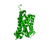 3cllC 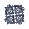 3cn6C 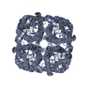 1z98S S: Starting model for refinement C: citing same article ( |
|---|---|
| Similar structure data |


-Links
 PDBj
PDBj
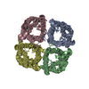
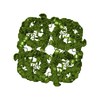
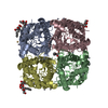
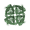
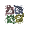
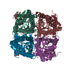
- Assembly
| Deposited unit | 
| |||||||||
|---|---|---|---|---|---|---|---|---|---|---|
| 1 | 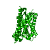
| |||||||||
| Unit cell |
| |||||||||
| Components on special symmetry positions |
|
-Components
| #1: Protein | Mass: 32642.828 Da / Num. of mol.: 1 / Mutation: S115E, S274E Source method: isolated from a genetically manipulated source Source: (gene. exp.) Spinacia oleracea (spinach) / Production host:  Pichia pastoris (fungus) / Strain (production host): X33 / References: UniProt: Q41372 Pichia pastoris (fungus) / Strain (production host): X33 / References: UniProt: Q41372 |
|---|---|
| #2: Water | ChemComp-HOH /  Mass: 18.015 Da / Num. of mol.: 114 / Source method: isolated from a natural source / Formula: H2O Mass: 18.015 Da / Num. of mol.: 114 / Source method: isolated from a natural source / Formula: H2O |
-Experimental details
-Experiment
| Experiment | Method: X-RAY DIFFRACTION / Number of used crystals: 1 |
|---|
- Sample preparation
| Crystal | Density Matthews: 2.53 Å3/Da / Density % sol: 51.48 % |
|---|---|
| Crystal grow | Temperature: 280 K / Method: vapor diffusion, hanging drop / pH: 6.5 Details: 30% PEG400, 0.1M NaCl, 0.1M MgCl2, 0.1M MES, pH 6.5, VAPOR DIFFUSION, HANGING DROP, temperature 280K |
-Data collection
| Diffraction | Mean temperature: 100 K |
|---|---|
| Diffraction source | Source: SYNCHROTRON / Site: ESRF  / Beamline: ID23-2 / Wavelength: 0.8726 Å / Beamline: ID23-2 / Wavelength: 0.8726 Å |
| Detector | Type: ADSC QUANTUM 4 / Detector: CCD / Date: Jul 2, 2007 |
| Radiation | Protocol: SINGLE WAVELENGTH / Monochromatic (M) / Laue (L): M / Scattering type: x-ray |
| Radiation wavelength | Wavelength: 0.8726 Å / Relative weight: 1 |
| Reflection | Resolution: 2.05→20 Å / Num. all: 20465 / Num. obs: 20465 / % possible obs: 100 % / Redundancy: 7.7 % / Rsym value: 0.084 |
| Reflection shell | Resolution: 2.05→2.16 Å / Redundancy: 7.6 % / Num. unique all: 3007 / Rsym value: 0.335 |
- Processing
| Software |
| ||||||||||||||||||||||||||||||||||||||||||||||||||||||||||||||||||||||||||||||||||||||||||
|---|---|---|---|---|---|---|---|---|---|---|---|---|---|---|---|---|---|---|---|---|---|---|---|---|---|---|---|---|---|---|---|---|---|---|---|---|---|---|---|---|---|---|---|---|---|---|---|---|---|---|---|---|---|---|---|---|---|---|---|---|---|---|---|---|---|---|---|---|---|---|---|---|---|---|---|---|---|---|---|---|---|---|---|---|---|---|---|---|---|---|---|
| Refinement | Method to determine structure: MOLECULAR REPLACEMENT Starting model: PDB entry 1Z98 Resolution: 2.05→20 Å / Cor.coef. Fo:Fc: 0.96 / Cor.coef. Fo:Fc free: 0.95 / SU B: 2.604 / SU ML: 0.074 / Cross valid method: THROUGHOUT / ESU R: 0.134 / ESU R Free: 0.119 / Stereochemistry target values: MAXIMUM LIKELIHOOD
| ||||||||||||||||||||||||||||||||||||||||||||||||||||||||||||||||||||||||||||||||||||||||||
| Solvent computation | Ion probe radii: 0.8 Å / Shrinkage radii: 0.8 Å / VDW probe radii: 1.2 Å / Solvent model: MASK | ||||||||||||||||||||||||||||||||||||||||||||||||||||||||||||||||||||||||||||||||||||||||||
| Displacement parameters | Biso mean: 26.465 Å2
| ||||||||||||||||||||||||||||||||||||||||||||||||||||||||||||||||||||||||||||||||||||||||||
| Refinement step | Cycle: LAST / Resolution: 2.05→20 Å
| ||||||||||||||||||||||||||||||||||||||||||||||||||||||||||||||||||||||||||||||||||||||||||
| Refine LS restraints |
| ||||||||||||||||||||||||||||||||||||||||||||||||||||||||||||||||||||||||||||||||||||||||||
| LS refinement shell | Resolution: 2.05→2.103 Å / Total num. of bins used: 20
|
