 Movie
Movie Controller

[English] 日本語
 Yorodumi
- PDB-2rgz: Ensemble refinement of the protein crystal structure of human hem... -
+ Open data

- Basic information
| Entry | Database: PDB / ID: 2rgz | ||||||
|---|---|---|---|---|---|---|---|
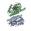
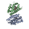
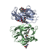
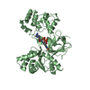
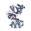
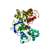
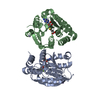
| Title | Ensemble refinement of the protein crystal structure of human heme oxygenase-2 C127A (HO-2) with bound heme | ||||||
 Components Components | Heme oxygenase 2 | ||||||
 Keywords Keywords | OXIDOREDUCTASE / Ensemble Refinement / Refinement Methodology Development / Structural Genomics Medical Relevance / Structural Genomics Community Request / HO-2 / HEME Oxygenase / Endoplasmic reticulum / Iron / Metal-binding / Microsome / Structural Genomics / Protein Structure Initiative / PSI / Center for Eukaryotic Structural Genomics / CESG | ||||||
| Function / homology |  Function and homology information Function and homology informationheme oxygenase (biliverdin-producing) / heme oxidation / heme oxygenase (decyclizing) activity / heme catabolic process / Heme degradation / RHOA GTPase cycle / specific granule membrane / Cytoprotection by HMOX1 / Iron uptake and transport / response to oxidative stress ...heme oxygenase (biliverdin-producing) / heme oxidation / heme oxygenase (decyclizing) activity / heme catabolic process / Heme degradation / RHOA GTPase cycle / specific granule membrane / Cytoprotection by HMOX1 / Iron uptake and transport / response to oxidative stress / response to hypoxia / heme binding / Neutrophil degranulation / endoplasmic reticulum membrane / membrane / metal ion binding / plasma membrane Similarity search - Function | ||||||
| Biological species |  Homo sapiens (human) Homo sapiens (human) | ||||||
| Method |  X-RAY DIFFRACTION / SYNCHROTRON / Multi-conformer re-refinement / Resolution: 2.61 Å X-RAY DIFFRACTION / SYNCHROTRON / Multi-conformer re-refinement / Resolution: 2.61 Å | ||||||
 Authors Authors | Bianchetti, C.M. / Bingman, C.A. / Bitto, E. / Wesenberg, G.E. / Phillips Jr., G.N. / Center for Eukaryotic Structural Genomics (CESG) | ||||||
 Citation Citation | Journal: J.Biol.Chem. / Year: 2007 Title: Comparison of Apo- and Heme-bound Crystal Structures of a Truncated Human Heme Oxygenase-2. Authors: Bianchetti, C.M. / Yi, L. / Ragsdale, S.W. / Phillips Jr., G.N. | ||||||
| History |
|
- Structure visualization
| Structure viewer | Molecule: MolmilJmol/JSmol |
|---|
- Downloads & links
-Download
| PDBx/mmCIF format | 2rgz.cif.gz | 1.3 MB | Display | PDBx/mmCIF format |
|---|---|---|---|---|
| PDB format | pdb2rgz.ent.gz | 1.1 MB | Display | PDB format |
| PDBx/mmJSON format | 2rgz.json.gz | Tree view | PDBx/mmJSON format | |
| Others |  Other downloads Other downloads |
-Validation report
| Arichive directory | https://data.pdbj.org/pub/pdb/validation_reports/rg/2rgzftp://data.pdbj.org/pub/pdb/validation_reports/rg/2rgz | HTTPS FTP |
|---|
-Related structure data
| Related structure data |  2q32C  2qppSC S: Starting model for refinement C: citing same article ( |
|---|---|
| Similar structure data | |
| Other databases |


-Links
 PDBj
PDBj


- Assembly
| Deposited unit | 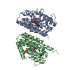
| ||||||||
|---|---|---|---|---|---|---|---|---|---|
| 1 |
| ||||||||
| 2 | 
| ||||||||
| 3 | 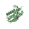
| ||||||||
| Unit cell |
| ||||||||
| Number of models | 16 |
-Components
| #1: Protein | Mass: 30491.271 Da / Num. of mol.: 2 / Mutation: C127A Source method: isolated from a genetically manipulated source Source: (gene. exp.) Homo sapiens (human) / Gene: HMOX2, HO2 / Plasmid: pGEX 4T-2 / Species (production host): Escherichia coli / Production host:  References: UniProt: P30519, heme oxygenase (biliverdin-producing) #2: Chemical |   Mass: 616.487 Da / Num. of mol.: 2 / Source method: obtained synthetically / Formula: C34H32FeN4O4 Mass: 616.487 Da / Num. of mol.: 2 / Source method: obtained synthetically / Formula: C34H32FeN4O4#3: Water | ChemComp-HOH / |  Mass: 18.015 Da / Num. of mol.: 83 / Source method: isolated from a natural source / Formula: H2O Mass: 18.015 Da / Num. of mol.: 83 / Source method: isolated from a natural source / Formula: H2O |
|---|
-Experimental details
-Experiment
| Experiment | Method: X-RAY DIFFRACTION / Number of used crystals: 1 |
|---|
- Sample preparation
| Crystal | Density Matthews: 2.56 Å3/Da / Density % sol: 51.94 % |
|---|---|
| Crystal grow | Temperature: 277 K / Method: vapor diffusion, hanging drop Details: Protein solution (5 mg/ml Protein, 1:1 ratio Heme, 2% DMSO, 0.050 M Potassium chloride, 0.050 M Tris-HCl pH 7.5) mixed in a 1.5:1 ratio with the Well solution (33% PEG DME 500, 0.020 M ...Details: Protein solution (5 mg/ml Protein, 1:1 ratio Heme, 2% DMSO, 0.050 M Potassium chloride, 0.050 M Tris-HCl pH 7.5) mixed in a 1.5:1 ratio with the Well solution (33% PEG DME 500, 0.020 M Magnesium chloride, 0.10 M HEPES pH 7.5). Cryoprotected with well solution, VAPOR DIFFUSION, HANGING DROP, temperature 277K |
-Data collection
| Diffraction | Mean temperature: 100 K | |||||||||||||||||||||||||||||||||||||||||||||||||||||||||||||||||||||||||||||
|---|---|---|---|---|---|---|---|---|---|---|---|---|---|---|---|---|---|---|---|---|---|---|---|---|---|---|---|---|---|---|---|---|---|---|---|---|---|---|---|---|---|---|---|---|---|---|---|---|---|---|---|---|---|---|---|---|---|---|---|---|---|---|---|---|---|---|---|---|---|---|---|---|---|---|---|---|---|---|
| Diffraction source | Source: SYNCHROTRON / Site: APS  / Beamline: 23-ID-B / Wavelength: 0.97946 Å / Beamline: 23-ID-B / Wavelength: 0.97946 Å | |||||||||||||||||||||||||||||||||||||||||||||||||||||||||||||||||||||||||||||
| Detector | Type: MARMOSAIC 300 mm CCD / Detector: CCD / Date: Jul 6, 2007 / Details: Adjustable focus K-B pair Si plus Pt, Rh coatings | |||||||||||||||||||||||||||||||||||||||||||||||||||||||||||||||||||||||||||||
| Radiation | Monochromator: Double crystal cryo-cooled Si(111) / Protocol: SINGLE WAVELENGTH / Monochromatic (M) / Laue (L): M / Scattering type: x-ray | |||||||||||||||||||||||||||||||||||||||||||||||||||||||||||||||||||||||||||||
| Radiation wavelength | Wavelength: 0.97946 Å / Relative weight: 1 | |||||||||||||||||||||||||||||||||||||||||||||||||||||||||||||||||||||||||||||
| Reflection | Resolution: 2.32→42.413 Å / Num. obs: 24030 / % possible obs: 85.6 % / Redundancy: 11.6 % / Rmerge(I) obs: 0.083 / Χ2: 1.039 / Net I/σ(I): 16.268 | |||||||||||||||||||||||||||||||||||||||||||||||||||||||||||||||||||||||||||||
| Reflection shell | Diffraction-ID: 1
|
- Processing
| Software |
| ||||||||||||||||||||||||||||||||||||||||||||||||||||||||||||||||||||||
|---|---|---|---|---|---|---|---|---|---|---|---|---|---|---|---|---|---|---|---|---|---|---|---|---|---|---|---|---|---|---|---|---|---|---|---|---|---|---|---|---|---|---|---|---|---|---|---|---|---|---|---|---|---|---|---|---|---|---|---|---|---|---|---|---|---|---|---|---|---|---|---|
| Refinement | Method to determine structure: Multi-conformer re-refinement Starting model: PDB entry 2QPP Resolution: 2.61→39.02 Å / Rfactor Rfree error: 0.008 / Data cutoff high absF: 1772722.5 / Data cutoff low absF: 0 / Isotropic thermal model: RESTRAINED / Cross valid method: THROUGHOUT / σ(F): 0 Stereochemistry target values: maximum likelihood using amplitudes Details: This PDB entry is a re-refinement using an ensemble model of the previously deposited single-conformer structure 2qpp and the first data set in the deposited structure factor file for 2qpp ...Details: This PDB entry is a re-refinement using an ensemble model of the previously deposited single-conformer structure 2qpp and the first data set in the deposited structure factor file for 2qpp along with the R-free set defined therein. The coordinates were generated by an automated protocol from an initial model consisting of 16 identical copies of the protein and non-water hetero-atoms assigned fractional occupancies adding up to one, and a single copy of the solvent molecules. Refinement was carried out with all the conformers present simultaneously and with the potential energy terms corresponding to interactions between the different conformers excluded. The helix and sheet records were calculated using coordinates from the first conformer only. The structure visualization program PYMOL is well-suited for directly viewing the ensemble model presented in this PDB file.
| ||||||||||||||||||||||||||||||||||||||||||||||||||||||||||||||||||||||
| Solvent computation | Solvent model: FLAT MODEL / Bsol: 53.313 Å2 / ksol: 0.329 e/Å3 | ||||||||||||||||||||||||||||||||||||||||||||||||||||||||||||||||||||||
| Displacement parameters | Biso mean: 56.5 Å2
| ||||||||||||||||||||||||||||||||||||||||||||||||||||||||||||||||||||||
| Refine analyze |
| ||||||||||||||||||||||||||||||||||||||||||||||||||||||||||||||||||||||
| Refinement step | Cycle: LAST / Resolution: 2.61→39.02 Å
| ||||||||||||||||||||||||||||||||||||||||||||||||||||||||||||||||||||||
| Refine LS restraints |
| ||||||||||||||||||||||||||||||||||||||||||||||||||||||||||||||||||||||
| LS refinement shell | Refine-ID: X-RAY DIFFRACTION / Total num. of bins used: 6
| ||||||||||||||||||||||||||||||||||||||||||||||||||||||||||||||||||||||
| Xplor file |
|
