 Movie
Movie Controller


+ Open data

- Basic information
| Entry | Database: PDB / ID: 1ddi | ||||||
|---|---|---|---|---|---|---|---|
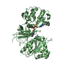
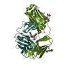
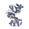
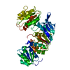
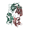
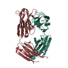
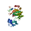
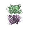
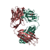
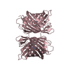
| Title | CRYSTAL STRUCTURE OF SIR-FP60 | ||||||
 Components Components | SULFITE REDUCTASE [NADPH] FLAVOPROTEIN ALPHA-COMPONENT | ||||||
 Keywords Keywords | OXIDOREDUCTASE / CYTOCHROME P450 REDUCTASE / FNR / FLAVOPROTEIN / MODULAR PROTEIN | ||||||
| Function / homology |  Function and homology information Function and homology informationassimilatory sulfite reductase (NADPH) / sulfite reductase (NADPH) activity / sulfite reductase complex (NADPH) / riboflavin reductase (NADPH) activity / sulfate assimilation / hydrogen sulfide biosynthetic process / L-cysteine biosynthetic process / NADP+ binding / FMN binding / flavin adenine dinucleotide binding ...assimilatory sulfite reductase (NADPH) / sulfite reductase (NADPH) activity / sulfite reductase complex (NADPH) / riboflavin reductase (NADPH) activity / sulfate assimilation / hydrogen sulfide biosynthetic process / L-cysteine biosynthetic process / NADP+ binding / FMN binding / flavin adenine dinucleotide binding / oxidoreductase activity / cytosol Similarity search - Function | ||||||
| Biological species |  | ||||||
| Method |  X-RAY DIFFRACTION / SYNCHROTRON / MIR / Resolution: 2.51 Å X-RAY DIFFRACTION / SYNCHROTRON / MIR / Resolution: 2.51 Å | ||||||
 Authors Authors | Gruez, A. / Pignol, D. / Zeghouf, M. / Coves, J. / Fontecave, M. / Ferrer, J.L. / Fontecilla-Camps, J.C. | ||||||
 Citation Citation | Journal: J.Mol.Biol. / Year: 2000 Title: Four crystal structures of the 60 kDa flavoprotein monomer of the sulfite reductase indicate a disordered flavodoxin-like module. Authors: Gruez, A. / Pignol, D. / Zeghouf, M. / Coves, J. / Fontecave, M. / Ferrer, J.L. / Fontecilla-Camps, J.C. | ||||||
| History |
|
- Structure visualization
| Structure viewer | Molecule: MolmilJmol/JSmol |
|---|
- Downloads & links
-Download
| PDBx/mmCIF format | 1ddi.cif.gz | 99.4 KB | Display | PDBx/mmCIF format |
|---|---|---|---|---|
| PDB format | pdb1ddi.ent.gz | 72.1 KB | Display | PDB format |
| PDBx/mmJSON format | 1ddi.json.gz | Tree view | PDBx/mmJSON format | |
| Others |  Other downloads Other downloads |
-Validation report
| Arichive directory | https://data.pdbj.org/pub/pdb/validation_reports/dd/1ddiftp://data.pdbj.org/pub/pdb/validation_reports/dd/1ddi | HTTPS FTP |
|---|
-Related structure data

-Links
 PDBj
PDBj

- Assembly
| Deposited unit | 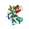
| ||||||||
|---|---|---|---|---|---|---|---|---|---|
| 1 |
| ||||||||
| Unit cell |
|
-Components
| #1: Protein | Mass: 42329.473 Da / Num. of mol.: 1 / Fragment: SIR-FP60 Source method: isolated from a genetically manipulated source Source: (gene. exp.) References: UniProt: P38038, assimilatory sulfite reductase (NADPH) |
|---|---|
| #2: Chemical | ChemComp-FAD /   Mass: 785.550 Da / Num. of mol.: 1 / Source method: obtained synthetically / Formula: C27H33N9O15P2 / Comment: FAD*YM Mass: 785.550 Da / Num. of mol.: 1 / Source method: obtained synthetically / Formula: C27H33N9O15P2 / Comment: FAD*YM |
| #3: Chemical | ChemComp-NAP /   Mass: 743.405 Da / Num. of mol.: 1 / Source method: obtained synthetically / Formula: C21H28N7O17P3 Mass: 743.405 Da / Num. of mol.: 1 / Source method: obtained synthetically / Formula: C21H28N7O17P3 |
| #4: Water | ChemComp-HOH /  Mass: 18.015 Da / Num. of mol.: 271 / Source method: isolated from a natural source / Formula: H2O Mass: 18.015 Da / Num. of mol.: 271 / Source method: isolated from a natural source / Formula: H2O |
-Experimental details
-Experiment
| Experiment | Method: X-RAY DIFFRACTION / Number of used crystals: 1 |
|---|
- Sample preparation
| Crystal | Density Matthews: 4.11 Å3/Da / Density % sol: 70.06 % | ||||||||||||||||||||||||||||||||||||
|---|---|---|---|---|---|---|---|---|---|---|---|---|---|---|---|---|---|---|---|---|---|---|---|---|---|---|---|---|---|---|---|---|---|---|---|---|---|
| Crystal grow | Temperature: 277 K / Method: vapor diffusion, hanging drop / pH: 7.5 Details: AMMONIUM SULFATE, pH 7.5, VAPOR DIFFUSION, HANGING DROP, temperature 4K | ||||||||||||||||||||||||||||||||||||
| Crystal grow | *PLUS pH: 7 | ||||||||||||||||||||||||||||||||||||
| Components of the solutions | *PLUS
|
-Data collection
| Diffraction | Mean temperature: 100 K |
|---|---|
| Diffraction source | Source: SYNCHROTRON / Site: ESRF  / Beamline: BM14 / Wavelength: 1 / Beamline: BM14 / Wavelength: 1 |
| Detector | Type: MARRESEARCH / Detector: IMAGE PLATE |
| Radiation | Protocol: SINGLE WAVELENGTH / Monochromatic (M) / Laue (L): M / Scattering type: x-ray |
| Radiation wavelength | Wavelength: 1 Å / Relative weight: 1 |
| Reflection | Resolution: 2.51→20 Å / Num. obs: 23679 / % possible obs: 98.4 % / Observed criterion σ(F): 0 / Observed criterion σ(I): 0 / Redundancy: 9 % / Rmerge(I) obs: 0.099 / Net I/σ(I): 6.2 |
| Reflection | *PLUS Num. obs: 23679 / Num. measured all: 212439 |
- Processing
| Software |
| ||||||||||||||||||||
|---|---|---|---|---|---|---|---|---|---|---|---|---|---|---|---|---|---|---|---|---|---|
| Refinement | Method to determine structure: MIR / Resolution: 2.51→20 Å / Cross valid method: FREE R-VALUE / σ(F): 0 / σ(I): 0
| ||||||||||||||||||||
| Refinement step | Cycle: LAST / Resolution: 2.51→20 Å
| ||||||||||||||||||||
| Software | *PLUS Name: REFMAC / Classification: refinement | ||||||||||||||||||||
| Refinement | *PLUS Lowest resolution: 20 Å / σ(F): 0 / Rfactor all: 0.1852 | ||||||||||||||||||||
| Solvent computation | *PLUS | ||||||||||||||||||||
| Displacement parameters | *PLUS | ||||||||||||||||||||
| Refine LS restraints | *PLUS
|
