 Movie
Movie Controller


+ Open data

- Basic information
| Entry | Database: PDB / ID: 1e0d | ||||||
|---|---|---|---|---|---|---|---|
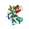
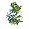
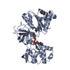
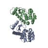
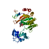
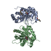
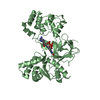
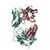
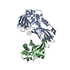
| Title | UDP-N-Acetylmuramoyl-L-Alanine:D-Glutamate Ligase | ||||||
 Components Components | UDP-N-ACETYLMURAMOYLALANINE--D-GLUTAMATE LIGASE | ||||||
 Keywords Keywords | LIGASE / PEPTIDOGLYCAN SYNTHESIS / MURD / ADP-FORMING ENZYME | ||||||
| Function / homology |  Function and homology information Function and homology informationUDP-N-acetylmuramoyl-L-alanine-D-glutamate ligase / UDP-N-acetylmuramoylalanine-D-glutamate ligase activity / peptidoglycan biosynthetic process / cell wall organization / regulation of cell shape / cell division / ATP binding / identical protein binding / cytoplasm Similarity search - Function | ||||||
| Biological species |  | ||||||
| Method |  X-RAY DIFFRACTION / SYNCHROTRON / MOLECULAR REPLACEMENT / Resolution: 2.4 Å X-RAY DIFFRACTION / SYNCHROTRON / MOLECULAR REPLACEMENT / Resolution: 2.4 Å | ||||||
 Authors Authors | Fanchon, E. / Bertrand, J. / Chantalat, L. / Dideberg, O. | ||||||
 Citation Citation | Journal: J.Mol.Biol. / Year: 2000 Title: "Open" Structures of Murd: Domain Movements and Structural Similarities with Folylpolyglutamate Synthetase. Authors: Bertrand, J. / Fanchon, E. / Martin, L. / Chantalat, L. / Auger, G. / Blanot, D. / Van Heijenoort, J. / Dideberg, O. | ||||||
| History |
|
- Structure visualization
| Structure viewer | Molecule: MolmilJmol/JSmol |
|---|
- Downloads & links
-Download
| PDBx/mmCIF format | 1e0d.cif.gz | 98.5 KB | Display | PDBx/mmCIF format |
|---|---|---|---|---|
| PDB format | pdb1e0d.ent.gz | 74 KB | Display | PDB format |
| PDBx/mmJSON format | 1e0d.json.gz | Tree view | PDBx/mmJSON format | |
| Others |  Other downloads Other downloads |
-Validation report
| Arichive directory | https://data.pdbj.org/pub/pdb/validation_reports/e0/1e0dftp://data.pdbj.org/pub/pdb/validation_reports/e0/1e0d | HTTPS FTP |
|---|
-Related structure data
| Related structure data | 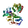 1eehC 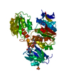 1uagS S: Starting model for refinement C: citing same article ( |
|---|---|
| Similar structure data |

-Links
 PDBj
PDBj- Assembly
| Deposited unit | 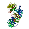
| ||||||||
|---|---|---|---|---|---|---|---|---|---|
| 1 |
| ||||||||
| Unit cell |
|
-Components
| #1: Protein | Mass: 46889.250 Da / Num. of mol.: 1 Source method: isolated from a genetically manipulated source Source: (gene. exp.) References: UniProt: P14900, UDP-N-acetylmuramoyl-L-alanine-D-glutamate ligase |
|---|---|
| #2: Chemical | ChemComp-SO4 /   Mass: 96.063 Da / Num. of mol.: 1 / Source method: obtained synthetically / Formula: SO4 Mass: 96.063 Da / Num. of mol.: 1 / Source method: obtained synthetically / Formula: SO4 |
| #3: Water | ChemComp-HOH /  Mass: 18.015 Da / Num. of mol.: 229 / Source method: isolated from a natural source / Formula: H2O Mass: 18.015 Da / Num. of mol.: 229 / Source method: isolated from a natural source / Formula: H2O |
-Experimental details
-Experiment
| Experiment | Method: X-RAY DIFFRACTION / Number of used crystals: 1 |
|---|
- Sample preparation
| Crystal | Density Matthews: 2.51 Å3/Da / Density % sol: 50.96 % | ||||||||||||||||||||||||||||||||||||||||||||||||
|---|---|---|---|---|---|---|---|---|---|---|---|---|---|---|---|---|---|---|---|---|---|---|---|---|---|---|---|---|---|---|---|---|---|---|---|---|---|---|---|---|---|---|---|---|---|---|---|---|---|
| Crystal grow | pH: 6 / Details: pH 6.00 | ||||||||||||||||||||||||||||||||||||||||||||||||
| Crystal grow | *PLUS Temperature: 15 ℃ / pH: 7.5 / Method: vapor diffusion, hanging dropDetails: drop solution was mixed with an equal volume of reservoir solution | ||||||||||||||||||||||||||||||||||||||||||||||||
| Components of the solutions | *PLUS
|
-Data collection
| Diffraction | Mean temperature: 100 K |
|---|---|
| Diffraction source | Source: SYNCHROTRON / Site: ESRF  / Beamline: D2AM / Wavelength: 0.9801 / Beamline: D2AM / Wavelength: 0.9801 |
| Detector | Type: MARRESEARCH / Detector: CCD / Date: Oct 23, 1997 / Details: MIRRORS |
| Radiation | Monochromator: DOUBLE CRYSTAL SI(111) / Protocol: SINGLE WAVELENGTH / Monochromatic (M) / Laue (L): M / Scattering type: x-ray |
| Radiation wavelength | Wavelength: 0.9801 Å / Relative weight: 1 |
| Reflection | Resolution: 2.4→50 Å / Num. obs: 19294 / % possible obs: 99 % / Redundancy: 7.3 % / Biso Wilson estimate: 25.5 Å2 / Rsym value: 0.04 / Net I/σ(I): 13 |
| Reflection shell | Resolution: 2.4→2.8 Å / Redundancy: 6.1 % / Mean I/σ(I) obs: 3.7 / Rsym value: 0.21 / % possible all: 99.9 |
| Reflection | *PLUS % possible obs: 98.9 % / Rmerge(I) obs: 0.088 |
| Reflection shell | *PLUS % possible obs: 99.9 % / Rmerge(I) obs: 0.212 |
- Processing
| Software |
| ||||||||||||||||||||||||||||||||||||||||||||||||||||||||||||||||||||||||||||||||
|---|---|---|---|---|---|---|---|---|---|---|---|---|---|---|---|---|---|---|---|---|---|---|---|---|---|---|---|---|---|---|---|---|---|---|---|---|---|---|---|---|---|---|---|---|---|---|---|---|---|---|---|---|---|---|---|---|---|---|---|---|---|---|---|---|---|---|---|---|---|---|---|---|---|---|---|---|---|---|---|---|---|
| Refinement | Method to determine structure: MOLECULAR REPLACEMENT Starting model: 2 DOMAINS FROM 1UAG Resolution: 2.4→20 Å / Rfactor Rfree error: 0.009 / Data cutoff high absF: 1911015.04 / Isotropic thermal model: RESTRAINED / Cross valid method: THROUGHOUT / σ(F): 0 Details: RESIDUES 183-188 AND 221 - 222 WERE NOT VISIBLE IN THE ELECTRON DENSITY AND THEIR COORDINATES ARE NOT INCLUDED IN THIS ENTRY. RESIDUES 183-188 AND 221 - 222 WERE NOT VISIBLE IN THE ELECTRON ...Details: RESIDUES 183-188 AND 221 - 222 WERE NOT VISIBLE IN THE ELECTRON DENSITY AND THEIR COORDINATES ARE NOT INCLUDED IN THIS ENTRY. RESIDUES 183-188 AND 221 - 222 WERE NOT VISIBLE IN THE ELECTRON DENSITY AND THEIR COORDINATES ARE NOT INCLUDED IN THIS ENTRY.
| ||||||||||||||||||||||||||||||||||||||||||||||||||||||||||||||||||||||||||||||||
| Solvent computation | Solvent model: FLAT MODEL / Bsol: 63.2219 Å2 / ksol: 0.365383 e/Å3 | ||||||||||||||||||||||||||||||||||||||||||||||||||||||||||||||||||||||||||||||||
| Displacement parameters | Biso mean: 40.5 Å2
| ||||||||||||||||||||||||||||||||||||||||||||||||||||||||||||||||||||||||||||||||
| Refine analyze |
| ||||||||||||||||||||||||||||||||||||||||||||||||||||||||||||||||||||||||||||||||
| Refinement step | Cycle: LAST / Resolution: 2.4→20 Å
| ||||||||||||||||||||||||||||||||||||||||||||||||||||||||||||||||||||||||||||||||
| Refine LS restraints |
| ||||||||||||||||||||||||||||||||||||||||||||||||||||||||||||||||||||||||||||||||
| LS refinement shell | Resolution: 2.4→2.55 Å / Rfactor Rfree error: 0.033 / Total num. of bins used: 6
| ||||||||||||||||||||||||||||||||||||||||||||||||||||||||||||||||||||||||||||||||
| Xplor file |
| ||||||||||||||||||||||||||||||||||||||||||||||||||||||||||||||||||||||||||||||||
| Software | *PLUS Name: CNS / Version: 0.4 / Classification: refinement | ||||||||||||||||||||||||||||||||||||||||||||||||||||||||||||||||||||||||||||||||
| Refinement | *PLUS | ||||||||||||||||||||||||||||||||||||||||||||||||||||||||||||||||||||||||||||||||
| Solvent computation | *PLUS | ||||||||||||||||||||||||||||||||||||||||||||||||||||||||||||||||||||||||||||||||
| Displacement parameters | *PLUS Biso mean: 40.7 Å2 | ||||||||||||||||||||||||||||||||||||||||||||||||||||||||||||||||||||||||||||||||
| Refine LS restraints | *PLUS
|
