 Movie
Movie Controller

[English] 日本語
 Yorodumi
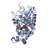
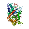
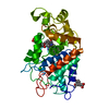
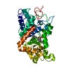
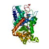
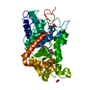
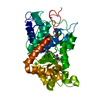
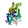
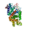
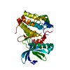
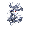
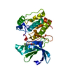
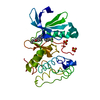
- PDB-1h5a: STRUCTURE OF FERRIC HORSERADISH PEROXIDASE C1A IN COMPLEX WITH ACETATE -
+ Open data

- Basic information
| Entry | Database: PDB / ID: 1h5a | ||||||
|---|---|---|---|---|---|---|---|
| Title | STRUCTURE OF FERRIC HORSERADISH PEROXIDASE C1A IN COMPLEX WITH ACETATE | ||||||
 Components Components | PEROXIDASE C1A | ||||||
 Keywords Keywords | OXIDOREDUCTASE / PEROXIDASE / HORSERADISH / FERRIC STATE / ACETATE ION | ||||||
| Function / homology |  Function and homology information Function and homology informationperoxidase / lactoperoxidase activity / vacuole / hydrogen peroxide catabolic process / response to oxidative stress / heme binding / extracellular region / metal ion binding Similarity search - Function | ||||||
| Biological species |  ARMORACIA RUSTICANA (horseradish) ARMORACIA RUSTICANA (horseradish) | ||||||
| Method |  X-RAY DIFFRACTION / SYNCHROTRON / OTHER / Resolution: 1.6 Å X-RAY DIFFRACTION / SYNCHROTRON / OTHER / Resolution: 1.6 Å | ||||||
 Authors Authors | Berglund, G.I. / Carlsson, G.H. / Hajdu, J. / Smith, A.T. / Szoke, H. / Henriksen, A. | ||||||
 Citation Citation | Journal: Nature / Year: 2002 Title: The Catalytic Pathway of Horseradish Peroxidase at High Resolution Authors: Berglund, G.I. / Carlsson, G.H. / Smith, A.T. / Szoke, H. / Henriksen, A. / Hajdu, J. #1: Journal: J.Biol.Chem. / Year: 1999Title: The Structures of the Horseradish Peroxidase C-Ferulic Acid Complex and the Ternary Complex with Cyanide Suggest How Peroxidases Oxidize Small Phenolic Substrates Authors: Henriksen, A. / Smith, A.T. / Gajhede, M. #2: Journal: Nat.Struct.Biol. / Year: 1997Title: Crystal Structure of Horseradish Peroxidase C at 2.15 A Resolution Authors: Gajhede, M. / Schuller, D.J. / Henriksen, A. / Smith, A.T. / Poulos, T.L. #3: Journal: J.Biol.Chem. / Year: 1990 Title: Expression of a Synthetic Gene for Horseradish Peroxidase C in Escherichia Coli and Folding and Activation of the Recombinant Enzyme with Ca2+ and Heme Authors: Smith, A.T. / Santama, N. / Dacey, S. / Edwards, M. / Bray, R.C. / Thorneley, R.N. / Burke, J.F. | ||||||
| History |
|
- Structure visualization
| Structure viewer | Molecule: MolmilJmol/JSmol |
|---|
- Downloads & links
-Download
| PDBx/mmCIF format | 1h5a.cif.gz | 86 KB | Display | PDBx/mmCIF format |
|---|---|---|---|---|
| PDB format | pdb1h5a.ent.gz | 63.8 KB | Display | PDB format |
| PDBx/mmJSON format | 1h5a.json.gz | Tree view | PDBx/mmJSON format | |
| Others |  Other downloads Other downloads |
-Validation report
| Summary document | 1h5a_validation.pdf.gz | 806.1 KB | Display | wwPDB validaton report |
|---|---|---|---|---|
| Full document | 1h5a_full_validation.pdf.gz | 807.3 KB | Display | |
| Data in XML | 1h5a_validation.xml.gz | 20.1 KB | Display | |
| Data in CIF | 1h5a_validation.cif.gz | 29.8 KB | Display | |
| Arichive directory | https://data.pdbj.org/pub/pdb/validation_reports/h5/1h5aftp://data.pdbj.org/pub/pdb/validation_reports/h5/1h5a | HTTPS FTP |
-Related structure data
| Related structure data | 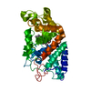 1h55C  1h57C  1h58C  1h5cC  1h5dC  1h5eC 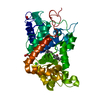 1h5fC 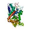 1h5gC  1h5hC  1h5iC 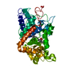 1h5jC  1h5kC  1h5lC  1h5mC 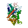 1hchC C: citing same article ( |
|---|---|
| Similar structure data |


-Links
 PDBj
PDBj
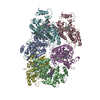
- Assembly
| Deposited unit | 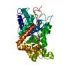
| ||||||||
|---|---|---|---|---|---|---|---|---|---|
| 1 |
| ||||||||
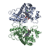
| Unit cell |
|
-Components
| #1: Protein | Mass: 33948.141 Da / Num. of mol.: 1 Source method: isolated from a genetically manipulated source Details: AN ACETATE ION IS BOUND IN THE ACTIVE SITE / Source: (gene. exp.) ARMORACIA RUSTICANA (horseradish) / Description: SYNTHETIC GENE / Production host:  | ||||||||
|---|---|---|---|---|---|---|---|---|---|
| #2: Chemical | ChemComp-HEM /   Mass: 616.487 Da / Num. of mol.: 1 / Source method: obtained synthetically / Formula: C34H32FeN4O4 Mass: 616.487 Da / Num. of mol.: 1 / Source method: obtained synthetically / Formula: C34H32FeN4O4 | ||||||||
| #3: Chemical |   Mass: 40.078 Da / Num. of mol.: 2 / Source method: obtained synthetically / Formula: Ca Mass: 40.078 Da / Num. of mol.: 2 / Source method: obtained synthetically / Formula: Ca#4: Chemical |   Mass: 59.044 Da / Num. of mol.: 2 / Source method: obtained synthetically / Formula: C2H3O2 Mass: 59.044 Da / Num. of mol.: 2 / Source method: obtained synthetically / Formula: C2H3O2#5: Water | ChemComp-HOH / |  Mass: 18.015 Da / Num. of mol.: 404 / Source method: isolated from a natural source / Formula: H2O Mass: 18.015 Da / Num. of mol.: 404 / Source method: isolated from a natural source / Formula: H2OHas protein modification | Y | Sequence details | THE SWS ENTRY INCLUDES N-TERM AND C-TERM SIGNAL PEPTIDES. | |
-Experimental details
-Experiment
| Experiment | Method: X-RAY DIFFRACTION / Number of used crystals: 7 |
|---|
- Sample preparation
| Crystal | Density Matthews: 2.25 Å3/Da / Density % sol: 45.3 % Description: STARTING MODEL FOR RIGID-BODY REFINEMENT WAS PDB ENTRY 7ATJ |
|---|---|
| Crystal grow | pH: 6.5 Details: 20% (W/V) PEG 4000, 0.2 M CALCIUM ACETATE, 0.1 M CACODYLATE BUFFER, PH 6.5 |
-Data collection
| Diffraction | Mean temperature: 100 K |
|---|---|
| Diffraction source | Source: SYNCHROTRON / Site: ESRF  / Beamline: ID14-3 / Wavelength: 0.931 / Beamline: ID14-3 / Wavelength: 0.931 |
| Detector | Type: MARRESEARCH / Detector: CCD / Date: Feb 15, 1999 / Details: MULTILAYER |
| Radiation | Monochromator: DIAMOND (111) / Protocol: SINGLE WAVELENGTH / Monochromatic (M) / Laue (L): M / Scattering type: x-ray |
| Radiation wavelength | Wavelength: 0.931 Å / Relative weight: 1 |
| Reflection | Resolution: 1.6→19.7 Å / Num. obs: 42167 / % possible obs: 97.7 % / Redundancy: 4.36 % / Biso Wilson estimate: 17.6 Å2 / Rmerge(I) obs: 0.058 / Net I/σ(I): 18.2 |
| Reflection shell | Resolution: 1.6→1.64 Å / Rmerge(I) obs: 0.203 / Mean I/σ(I) obs: 6.8 / % possible all: 93.9 |
- Processing
| Software |
| ||||||||||||||||||||||||||||||||||||||||||||||||||||||||||||||||||||||||||||||||
|---|---|---|---|---|---|---|---|---|---|---|---|---|---|---|---|---|---|---|---|---|---|---|---|---|---|---|---|---|---|---|---|---|---|---|---|---|---|---|---|---|---|---|---|---|---|---|---|---|---|---|---|---|---|---|---|---|---|---|---|---|---|---|---|---|---|---|---|---|---|---|---|---|---|---|---|---|---|---|---|---|---|
| Refinement | Method to determine structure: OTHER / Resolution: 1.6→19.71 Å / Rfactor Rfree error: 0.004 / Data cutoff high absF: 1344685.61 / Data cutoff low absF: 0 / Isotropic thermal model: RESTRAINED / Cross valid method: THROUGHOUT / σ(F): 0 Details: SER 306 WAS THE LAST RESIDUE SEEN IN THE ELECTRON DENSITY MAP THE FOLLOWING RESIDUES HAVE BEEN MODELLED IN DUAL CONFORMATIONS: 15,24,91,151,161,188,219,240, 249,286,296
| ||||||||||||||||||||||||||||||||||||||||||||||||||||||||||||||||||||||||||||||||
| Solvent computation | Solvent model: FLAT MODEL / Bsol: 49.644 Å2 / ksol: 0.358411 e/Å3 | ||||||||||||||||||||||||||||||||||||||||||||||||||||||||||||||||||||||||||||||||
| Displacement parameters | Biso mean: 16.7 Å2
| ||||||||||||||||||||||||||||||||||||||||||||||||||||||||||||||||||||||||||||||||
| Refine analyze |
| ||||||||||||||||||||||||||||||||||||||||||||||||||||||||||||||||||||||||||||||||
| Refinement step | Cycle: LAST / Resolution: 1.6→19.71 Å
| ||||||||||||||||||||||||||||||||||||||||||||||||||||||||||||||||||||||||||||||||
| Refine LS restraints |
| ||||||||||||||||||||||||||||||||||||||||||||||||||||||||||||||||||||||||||||||||
| LS refinement shell | Resolution: 1.6→1.7 Å / Rfactor Rfree error: 0.014 / Total num. of bins used: 6
| ||||||||||||||||||||||||||||||||||||||||||||||||||||||||||||||||||||||||||||||||
| Xplor file |
|
