 Movie
Movie Controller


+ Open data

- Basic information
| Entry | Database: PDB / ID: 1atj | ||||||
|---|---|---|---|---|---|---|---|
| Title | RECOMBINANT HORSERADISH PEROXIDASE C1A | ||||||
 Components Components | PEROXIDASE C1A | ||||||
 Keywords Keywords | OXIDOREDUCTASE / PEROXIDASE / GLYCOPROTEIN | ||||||
| Function / homology |  Function and homology information Function and homology informationlactoperoxidase activity / peroxidase / vacuole / hydrogen peroxide catabolic process / response to oxidative stress / heme binding / extracellular region / metal ion binding Similarity search - Function | ||||||
| Biological species |  Armoracia rusticana (horseradish) Armoracia rusticana (horseradish) | ||||||
| Method |  X-RAY DIFFRACTION / MOLECULAR REPLACEMENT / Resolution: 2.15 Å X-RAY DIFFRACTION / MOLECULAR REPLACEMENT / Resolution: 2.15 Å | ||||||
 Authors Authors | Gajhede, M. / Schuller, D.J. / Henriksen, A. / Smith, A.T. / Poulos, T.L. | ||||||
 Citation Citation | Journal: Nat.Struct.Biol. / Year: 1997 Title: Crystal structure of horseradish peroxidase C at 2.15 A resolution. Authors: Gajhede, M. / Schuller, D.J. / Henriksen, A. / Smith, A.T. / Poulos, T.L. #1: Journal: Acta Crystallogr.,Sect.D / Year: 1995Title: Crystallization and Preliminary X-Ray Studies of Recombinant Horseradish Peroxidase Authors: Henriksen, A. / Gajhede, M. / Baker, P. / Smith, A.T. / Burke, J.F. | ||||||
| History |
|
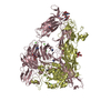
- Structure visualization
| Structure viewer | Molecule: MolmilJmol/JSmol |
|---|
- Downloads & links
-Download
| PDBx/mmCIF format | 1atj.cif.gz | 445.3 KB | Display | PDBx/mmCIF format |
|---|---|---|---|---|
| PDB format | pdb1atj.ent.gz | 365.2 KB | Display | PDB format |
| PDBx/mmJSON format | 1atj.json.gz | Tree view | PDBx/mmJSON format | |
| Others |  Other downloads Other downloads |
-Validation report
| Arichive directory | https://data.pdbj.org/pub/pdb/validation_reports/at/1atjftp://data.pdbj.org/pub/pdb/validation_reports/at/1atj | HTTPS FTP |
|---|
-Related structure data
| Related structure data |  1schS S: Starting model for refinement |
|---|---|
| Similar structure data |


-Links
 PDBj
PDBj
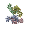
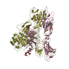
- Assembly
| Deposited unit | 
| ||||||||||||||||||||||||
|---|---|---|---|---|---|---|---|---|---|---|---|---|---|---|---|---|---|---|---|---|---|---|---|---|---|
| 1 |
| ||||||||||||||||||||||||
| Unit cell |
| ||||||||||||||||||||||||
| Noncrystallographic symmetry (NCS) | NCS oper:
|
-Components
| #1: Protein | Mass: 33746.961 Da / Num. of mol.: 6 Source method: isolated from a genetically manipulated source Source: (gene. exp.) Armoracia rusticana (horseradish) / Production host:  #2: Chemical | ChemComp-CA /   Mass: 40.078 Da / Num. of mol.: 12 / Source method: obtained synthetically / Formula: Ca Mass: 40.078 Da / Num. of mol.: 12 / Source method: obtained synthetically / Formula: Ca#3: Chemical | ChemComp-HEM /   Mass: 616.487 Da / Num. of mol.: 6 / Source method: obtained synthetically / Formula: C34H32FeN4O4 Mass: 616.487 Da / Num. of mol.: 6 / Source method: obtained synthetically / Formula: C34H32FeN4O4#4: Water | ChemComp-HOH / |  Mass: 18.015 Da / Num. of mol.: 888 / Source method: isolated from a natural source / Formula: H2O Mass: 18.015 Da / Num. of mol.: 888 / Source method: isolated from a natural source / Formula: H2OHas protein modification | Y | |
|---|
-Experimental details
-Experiment
| Experiment | Method: X-RAY DIFFRACTION / Number of used crystals: 1 |
|---|
- Sample preparation
| Crystal | Density Matthews: 2.04 Å3/Da / Density % sol: 39 % | ||||||||||||||||||||
|---|---|---|---|---|---|---|---|---|---|---|---|---|---|---|---|---|---|---|---|---|---|
| Crystal grow | pH: 6.5 Details: 16% (W/V) PEG 4000, 0.2 M ZINC ACETATE AND 0.1 M CACODYLATE BUFFER, PH 6.5 | ||||||||||||||||||||
| Crystal grow | *PLUS Method: vapor diffusion / Details: Braithwaite, A., (1976) J. Mol. Biol., 106, 229. | ||||||||||||||||||||
| Components of the solutions | *PLUS
|
-Data collection
| Diffraction | Mean temperature: 287 K |
|---|---|
| Diffraction source | Source: ROTATING ANODE / Type: RIGAKU RUH2R / Wavelength: 1.5418 |
| Detector | Type: RIGAKU / Detector: IMAGE PLATE / Date: Aug 1, 1995 |
| Radiation | Monochromator: GRAPHITE(002) / Monochromatic (M) / Laue (L): M / Scattering type: x-ray |
| Radiation wavelength | Wavelength: 1.5418 Å / Relative weight: 1 |
| Reflection | Resolution: 2.15→100 Å / Num. obs: 73216 / % possible obs: 83.6 % / Observed criterion σ(I): 0 / Redundancy: 1.7 % / Biso Wilson estimate: 12.8 Å2 / Rmerge(I) obs: 0.07 / Rsym value: 0.07 |
| Reflection shell | Resolution: 2.15→2.9 Å / Redundancy: 1.2 % / Rmerge(I) obs: 0.4 / Rsym value: 0.4 / % possible all: 65.4 |
| Reflection | *PLUS Num. measured all: 142833 |
| Reflection shell | *PLUS Lowest resolution: 2.19 Å / % possible obs: 65.4 % |
- Processing
| Software |
| ||||||||||||||||||||||||||||||||||||||||||||||||||||||||||||||||||||||||||||||||
|---|---|---|---|---|---|---|---|---|---|---|---|---|---|---|---|---|---|---|---|---|---|---|---|---|---|---|---|---|---|---|---|---|---|---|---|---|---|---|---|---|---|---|---|---|---|---|---|---|---|---|---|---|---|---|---|---|---|---|---|---|---|---|---|---|---|---|---|---|---|---|---|---|---|---|---|---|---|---|---|---|---|
| Refinement | Method to determine structure: MOLECULAR REPLACEMENT Starting model: PDB ENTRY 1SCH Resolution: 2.15→100 Å / Rfactor Rfree error: 0.003 / Data cutoff high absF: 10000000 / Data cutoff low absF: 0.001 / Isotropic thermal model: RESTRAINED / Cross valid method: THROUGHOUT / σ(F): 0 / Details: BULK SOLVENT MODEL USED
| ||||||||||||||||||||||||||||||||||||||||||||||||||||||||||||||||||||||||||||||||
| Displacement parameters | Biso mean: 24.9 Å2 | ||||||||||||||||||||||||||||||||||||||||||||||||||||||||||||||||||||||||||||||||
| Refine analyze |
| ||||||||||||||||||||||||||||||||||||||||||||||||||||||||||||||||||||||||||||||||
| Refinement step | Cycle: LAST / Resolution: 2.15→100 Å
| ||||||||||||||||||||||||||||||||||||||||||||||||||||||||||||||||||||||||||||||||
| Refine LS restraints |
| ||||||||||||||||||||||||||||||||||||||||||||||||||||||||||||||||||||||||||||||||
| Refine LS restraints NCS | NCS model details: CONSTR | ||||||||||||||||||||||||||||||||||||||||||||||||||||||||||||||||||||||||||||||||
| LS refinement shell | Resolution: 2.15→2.23 Å / Rfactor Rfree error: 0.013 / Total num. of bins used: 10
| ||||||||||||||||||||||||||||||||||||||||||||||||||||||||||||||||||||||||||||||||
| Xplor file |
| ||||||||||||||||||||||||||||||||||||||||||||||||||||||||||||||||||||||||||||||||
| Software | *PLUS Name: X-PLOR / Version: 3.851 / Classification: refinement | ||||||||||||||||||||||||||||||||||||||||||||||||||||||||||||||||||||||||||||||||
| Refine LS restraints | *PLUS
|
