 Movie
Movie Controller


+ Open data

- Basic information
| Entry | Database: PDB / ID: 6atj | ||||||
|---|---|---|---|---|---|---|---|
| Title | RECOMBINANT HORSERADISH PEROXIDASE C COMPLEX WITH FERULIC ACID | ||||||
 Components Components | PEROXIDASE C1A | ||||||
 Keywords Keywords | OXIDOREDUCTASE / PEROXIDASE / REDUCING SUBSTRATE | ||||||
| Function / homology |  Function and homology information Function and homology informationlactoperoxidase activity / peroxidase / vacuole / hydrogen peroxide catabolic process / response to oxidative stress / heme binding / extracellular region / metal ion binding Similarity search - Function | ||||||
| Biological species |  Armoracia rusticana (horseradish) Armoracia rusticana (horseradish) | ||||||
| Method |  X-RAY DIFFRACTION / MOLECULAR REPLACEMENT / Resolution: 2 Å X-RAY DIFFRACTION / MOLECULAR REPLACEMENT / Resolution: 2 Å | ||||||
 Authors Authors | Henriksen, A. / Smith, A.T. / Gajhede, M. | ||||||
 Citation Citation | Journal: J.Biol.Chem. / Year: 1999 Title: The structures of the horseradish peroxidase C-ferulic acid complex and the ternary complex with cyanide suggest how peroxidases oxidize small phenolic substrates. Authors: Henriksen, A. / Smith, A.T. / Gajhede, M. #1: Journal: Biochemistry / Year: 1998Title: Structural interactions between horseradish peroxidase C and the substrate benzhydroxamic acid determined by X-ray crystallography. Authors: Henriksen, A. / Schuller, D.J. / Meno, K. / Welinder, K.G. / Smith, A.T. / Gajhede, M. #2: Journal: Nat.Struct.Biol. / Year: 1997Title: Crystal structure of horseradish peroxidase C at 2.15 A resolution. Authors: Gajhede, M. / Schuller, D.J. / Henriksen, A. / Smith, A.T. / Poulos, T.L. #3: Journal: Acta Crystallogr.,Sect.D / Year: 1995 Title: Crystallization and preliminary X-ray studies of recombinant horseradish peroxidase. Authors: Henriksen, A. / Gajhede, M. / Baker, P. / Smith, A.T. / Burke, J.F. | ||||||
| History |
|
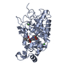
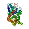
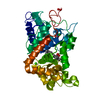
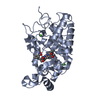
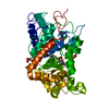
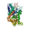
- Structure visualization
| Structure viewer | Molecule: MolmilJmol/JSmol |
|---|
- Downloads & links
-Download
| PDBx/mmCIF format | 6atj.cif.gz | 86.7 KB | Display | PDBx/mmCIF format |
|---|---|---|---|---|
| PDB format | pdb6atj.ent.gz | 64 KB | Display | PDB format |
| PDBx/mmJSON format | 6atj.json.gz | Tree view | PDBx/mmJSON format | |
| Others |  Other downloads Other downloads |
-Validation report
| Arichive directory | https://data.pdbj.org/pub/pdb/validation_reports/at/6atjftp://data.pdbj.org/pub/pdb/validation_reports/at/6atj | HTTPS FTP |
|---|
-Related structure data
| Related structure data | 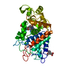 7atjC 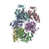 1atjS C: citing same article ( S: Starting model for refinement |
|---|---|
| Similar structure data |


-Links
 PDBj
PDBj
- Assembly
| Deposited unit | 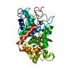
| ||||||||
|---|---|---|---|---|---|---|---|---|---|
| 1 |
| ||||||||
| Unit cell |
|
-Components
| #1: Protein | Mass: 33948.141 Da / Num. of mol.: 1 Source method: isolated from a genetically manipulated source Source: (gene. exp.) Armoracia rusticana (horseradish) / Description: SYNTHETIC GENE / Production host:  | ||||||||||
|---|---|---|---|---|---|---|---|---|---|---|---|
| #2: Chemical |   Mass: 40.078 Da / Num. of mol.: 2 / Source method: obtained synthetically / Formula: Ca Mass: 40.078 Da / Num. of mol.: 2 / Source method: obtained synthetically / Formula: Ca#3: Chemical | ChemComp-HEM / |   Mass: 616.487 Da / Num. of mol.: 1 / Source method: obtained synthetically / Formula: C34H32FeN4O4 Mass: 616.487 Da / Num. of mol.: 1 / Source method: obtained synthetically / Formula: C34H32FeN4O4#4: Chemical | ChemComp-FER / | 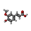  Mass: 194.184 Da / Num. of mol.: 1 / Source method: obtained synthetically / Formula: C10H10O4 Mass: 194.184 Da / Num. of mol.: 1 / Source method: obtained synthetically / Formula: C10H10O4#5: Water | ChemComp-HOH / |  Mass: 18.015 Da / Num. of mol.: 399 / Source method: isolated from a natural source / Formula: H2O Mass: 18.015 Da / Num. of mol.: 399 / Source method: isolated from a natural source / Formula: H2OHas protein modification | Y | Sequence details | THE SWS ENTRY INCLUDES N-TERM AND C-TERM SIGNAL PEPTIDES. | |
-Experimental details
-Experiment
| Experiment | Method: X-RAY DIFFRACTION / Number of used crystals: 1 |
|---|
- Sample preparation
| Crystal | Density Matthews: 2.35 Å3/Da / Density % sol: 47.5 % | ||||||||||||||||||||||||
|---|---|---|---|---|---|---|---|---|---|---|---|---|---|---|---|---|---|---|---|---|---|---|---|---|---|
| Crystal grow | pH: 6.5 Details: 16% (W/V) PEG 4000, 0.2 M CALCIUM ACETATE AND 0.1 M CACODYLATE BUFFER, PH 6.5 | ||||||||||||||||||||||||
| Crystal grow | *PLUS Temperature: 10 ℃ / Method: vapor diffusion | ||||||||||||||||||||||||
| Components of the solutions | *PLUS
|
-Data collection
| Diffraction | Mean temperature: 120 K |
|---|---|
| Diffraction source | Source: ROTATING ANODE / Type: RIGAKU RU200 / Wavelength: 1.5418 |
| Detector | Type: RIGAKU / Detector: IMAGE PLATE / Date: Jul 1, 1995 |
| Radiation | Monochromator: GRAPHITE / Protocol: SINGLE WAVELENGTH / Monochromatic (M) / Laue (L): M / Scattering type: x-ray |
| Radiation wavelength | Wavelength: 1.5418 Å / Relative weight: 1 |
| Reflection | Resolution: 2→29 Å / Num. obs: 21778 / % possible obs: 95.1 % / Observed criterion σ(I): 0 / Redundancy: 5 % / Biso Wilson estimate: 2.9 Å2 / Rmerge(I) obs: 0.076 |
| Reflection shell | Resolution: 2→2.07 Å / Rmerge(I) obs: 0.208 / % possible all: 96 |
| Reflection | *PLUS % possible obs: 97.2 % / Num. measured all: 108409 |
| Reflection shell | *PLUS % possible obs: 96 % |
- Processing
| Software |
| ||||||||||||||||||||||||||||||||||||||||||||||||||||||||||||
|---|---|---|---|---|---|---|---|---|---|---|---|---|---|---|---|---|---|---|---|---|---|---|---|---|---|---|---|---|---|---|---|---|---|---|---|---|---|---|---|---|---|---|---|---|---|---|---|---|---|---|---|---|---|---|---|---|---|---|---|---|---|
| Refinement | Method to determine structure: MOLECULAR REPLACEMENT Starting model: PDB ENTRY 1ATJ Resolution: 2→29 Å / Rfactor Rfree error: 0.006 / Data cutoff high rms absF: 344924.86 / Isotropic thermal model: RESTRAINED / Cross valid method: THROUGHOUT / σ(F): 0 / Details: BULK SOLVENT MODEL USED
| ||||||||||||||||||||||||||||||||||||||||||||||||||||||||||||
| Solvent computation | Solvent model: FLAT MODEL / Bsol: 57 Å2 / ksol: 0.349 e/Å3 | ||||||||||||||||||||||||||||||||||||||||||||||||||||||||||||
| Displacement parameters | Biso mean: 11.7 Å2
| ||||||||||||||||||||||||||||||||||||||||||||||||||||||||||||
| Refine analyze |
| ||||||||||||||||||||||||||||||||||||||||||||||||||||||||||||
| Refinement step | Cycle: LAST / Resolution: 2→29 Å
| ||||||||||||||||||||||||||||||||||||||||||||||||||||||||||||
| Refine LS restraints |
| ||||||||||||||||||||||||||||||||||||||||||||||||||||||||||||
| LS refinement shell | Resolution: 2→2.13 Å / Rfactor Rfree error: 0.019 / Total num. of bins used: 6
| ||||||||||||||||||||||||||||||||||||||||||||||||||||||||||||
| Software | *PLUS Name: CNS / Version: 0.5 / Classification: refinement | ||||||||||||||||||||||||||||||||||||||||||||||||||||||||||||
| Refinement | *PLUS Num. reflection obs: 21751 | ||||||||||||||||||||||||||||||||||||||||||||||||||||||||||||
| Solvent computation | *PLUS | ||||||||||||||||||||||||||||||||||||||||||||||||||||||||||||
| Displacement parameters | *PLUS | ||||||||||||||||||||||||||||||||||||||||||||||||||||||||||||
| Refine LS restraints | *PLUS
|
