 Movie
Movie Controller

[English] 日本語
 Yorodumi
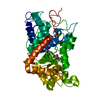
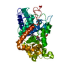
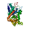
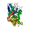
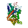
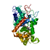
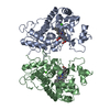
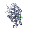
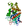
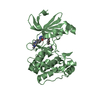
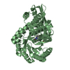
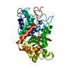
- PDB-1gx2: Recombinant horseradish peroxidase Phe209Ser complex with benzhyd... -
+ Open data

- Basic information
| Entry | Database: PDB / ID: 1gx2 | ||||||
|---|---|---|---|---|---|---|---|
| Title | Recombinant horseradish peroxidase Phe209Ser complex with benzhydroxamic acid | ||||||
 Components Components | PEROXIDASE C1A | ||||||
 Keywords Keywords | OXIDOREDUCTASE / PEROXIDASE | ||||||
| Function / homology |  Function and homology information Function and homology informationlactoperoxidase activity / peroxidase / vacuole / hydrogen peroxide catabolic process / response to oxidative stress / heme binding / extracellular region / metal ion binding Similarity search - Function | ||||||
| Biological species |  ARMORACIA RUSTICANA (horseradish) ARMORACIA RUSTICANA (horseradish) | ||||||
| Method |  X-RAY DIFFRACTION / MOLECULAR REPLACEMENT / Resolution: 2.2 Å X-RAY DIFFRACTION / MOLECULAR REPLACEMENT / Resolution: 2.2 Å | ||||||
 Authors Authors | Meno, K. / Gajhede, M. / Henriksen, A. | ||||||
 Citation Citation | Journal: To be Published Title: Hrpc Heme Crevice Architecture Authors: Henriksen, A. / Brissett, N. / Meno, K. / Mirza, O. / Gajhede, M. #1: Journal: J.Biol.Chem. / Year: 1999Title: The Structures of the Horseradish Peroxidase C-Ferulic Acid Complex and the Ternary Complex with Cyanide Suggest How Peroxidases Oxidize Small Phenolic Substrates Authors: Henriksen, A. / Smith, A.T. / Gajhede, M. #2: Journal: Biochemistry / Year: 1998Title: Structural Interactions between Horseradish Peroxidase C and the Substrate Benzhydroxamic Acid Determined by X-Ray Crystallography Authors: Henriksen, A. / Schuller, D.J. / Meno, K. / Welinder, K.G. / Smith, A.T. / Gajhede, M. #3: Journal: Nat.Struct.Biol. / Year: 1997Title: Crystal Structure of Horseradish Peroxidase C at 2.15 A Resolution Authors: Gajhede, M. / Schuller, D.J. / Henriksen, A. / Smith, A.T. / Poulos, T.L. | ||||||
| History |
|
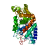
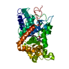
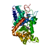
- Structure visualization
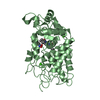
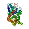
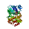
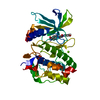
| Structure viewer | Molecule: MolmilJmol/JSmol |
|---|
- Downloads & links
-Download
| PDBx/mmCIF format | 1gx2.cif.gz | 141.4 KB | Display | PDBx/mmCIF format |
|---|---|---|---|---|
| PDB format | pdb1gx2.ent.gz | 110.4 KB | Display | PDB format |
| PDBx/mmJSON format | 1gx2.json.gz | Tree view | PDBx/mmJSON format | |
| Others |  Other downloads Other downloads |
-Validation report
| Arichive directory | https://data.pdbj.org/pub/pdb/validation_reports/gx/1gx2ftp://data.pdbj.org/pub/pdb/validation_reports/gx/1gx2 | HTTPS FTP |
|---|
-Related structure data
| Related structure data | 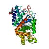 1gw2C 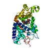 1gwoC 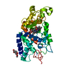 1gwtC  1gwuC 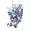 2atjS C: citing same article ( S: Starting model for refinement |
|---|---|
| Similar structure data |


-Links
 PDBj
PDBj
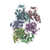
- Assembly
| Deposited unit | 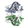
| ||||||||
|---|---|---|---|---|---|---|---|---|---|
| 1 | 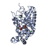
| ||||||||
| 2 | 
| ||||||||
| Unit cell |
|
-Components
| #1: Protein | Mass: 34019.246 Da / Num. of mol.: 2 / Mutation: YES Source method: isolated from a genetically manipulated source Source: (gene. exp.) ARMORACIA RUSTICANA (horseradish) / Description: SYNTHETIC GENE / Production host:  #2: Chemical |   Mass: 616.487 Da / Num. of mol.: 2 / Source method: obtained synthetically / Formula: C34H32FeN4O4 Mass: 616.487 Da / Num. of mol.: 2 / Source method: obtained synthetically / Formula: C34H32FeN4O4#3: Chemical | 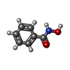  Mass: 137.136 Da / Num. of mol.: 2 / Source method: obtained synthetically / Formula: C7H7NO2 Mass: 137.136 Da / Num. of mol.: 2 / Source method: obtained synthetically / Formula: C7H7NO2#4: Chemical | ChemComp-CA /   Mass: 40.078 Da / Num. of mol.: 4 / Source method: obtained synthetically / Formula: Ca Mass: 40.078 Da / Num. of mol.: 4 / Source method: obtained synthetically / Formula: Ca#5: Water | ChemComp-HOH / |  Mass: 18.015 Da / Num. of mol.: 372 / Source method: isolated from a natural source / Formula: H2O Mass: 18.015 Da / Num. of mol.: 372 / Source method: isolated from a natural source / Formula: H2OCompound details | ENGINEERED | Has protein modification | Y | |
|---|
-Experimental details
-Experiment
| Experiment | Method: X-RAY DIFFRACTION / Number of used crystals: 1 |
|---|
- Sample preparation
| Crystal | Density Matthews: 2.6 Å3/Da / Density % sol: 52.78 % |
|---|---|
| Crystal grow | pH: 6.5 / Details: pH 6.50 |
-Data collection
| Diffraction | Mean temperature: 120 K |
|---|---|
| Diffraction source | Source: ROTATING ANODE / Type: RIGAKU RU200 / Wavelength: 1.5418 |
| Detector | Type: RIGAKU IMAGE PLATE / Detector: IMAGE PLATE / Date: Jun 15, 1997 |
| Radiation | Monochromator: GRAPHITE / Protocol: SINGLE WAVELENGTH / Monochromatic (M) / Laue (L): M / Scattering type: x-ray |
| Radiation wavelength | Wavelength: 1.5418 Å / Relative weight: 1 |
| Reflection | Resolution: 2.2→37.92 Å / Num. obs: 113464 / % possible obs: 96.6 % / Observed criterion σ(I): 0 / Redundancy: 3.3 % / Biso Wilson estimate: 4.7 Å2 / Rmerge(I) obs: 0.106 |
| Reflection shell | Resolution: 2.2→2.24 Å / Rmerge(I) obs: 0.391 / % possible all: 93.5 |
- Processing
| Software |
| ||||||||||||||||||||||||||||||||||||||||||||||||||||||||||||||||||||||||||||||||
|---|---|---|---|---|---|---|---|---|---|---|---|---|---|---|---|---|---|---|---|---|---|---|---|---|---|---|---|---|---|---|---|---|---|---|---|---|---|---|---|---|---|---|---|---|---|---|---|---|---|---|---|---|---|---|---|---|---|---|---|---|---|---|---|---|---|---|---|---|---|---|---|---|---|---|---|---|---|---|---|---|---|
| Refinement | Method to determine structure: MOLECULAR REPLACEMENT Starting model: PDB ENTRY 2ATJ Resolution: 2.2→37.92 Å / Rfactor Rfree error: 0.003 / Data cutoff high absF: 12108 / Isotropic thermal model: RESTRAINED / Cross valid method: THROUGHOUT / σ(F): 0 / Stereochemistry target values: MLF
| ||||||||||||||||||||||||||||||||||||||||||||||||||||||||||||||||||||||||||||||||
| Solvent computation | Solvent model: FLAT MODEL / Bsol: 47.4833 Å2 / ksol: 0.339209 e/Å3 | ||||||||||||||||||||||||||||||||||||||||||||||||||||||||||||||||||||||||||||||||
| Displacement parameters | Biso mean: 16.5 Å2
| ||||||||||||||||||||||||||||||||||||||||||||||||||||||||||||||||||||||||||||||||
| Refine analyze |
| ||||||||||||||||||||||||||||||||||||||||||||||||||||||||||||||||||||||||||||||||
| Refinement step | Cycle: LAST / Resolution: 2.2→37.92 Å
| ||||||||||||||||||||||||||||||||||||||||||||||||||||||||||||||||||||||||||||||||
| Refine LS restraints |
| ||||||||||||||||||||||||||||||||||||||||||||||||||||||||||||||||||||||||||||||||
| Refine LS restraints NCS | NCS model details: CONSTR | ||||||||||||||||||||||||||||||||||||||||||||||||||||||||||||||||||||||||||||||||
| LS refinement shell | Resolution: 2.2→2.34 Å / Rfactor Rfree error: 0.011 / Total num. of bins used: 6
|
