 Movie
Movie Controller

[English] 日本語
 Yorodumi
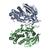
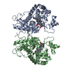
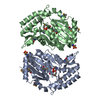
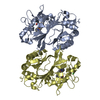
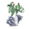
- PDB-3atj: HEME LIGAND MUTANT OF RECOMBINANT HORSERADISH PEROXIDASE IN COMPL... -
+ Open data

- Basic information
| Entry | Database: PDB / ID: 3atj | ||||||
|---|---|---|---|---|---|---|---|
| Title | HEME LIGAND MUTANT OF RECOMBINANT HORSERADISH PEROXIDASE IN COMPLEX WITH BENZHYDROXAMIC ACID | ||||||
 Components Components | PROTEIN (HORSERADISH PEROXIDASE C1A) | ||||||
 Keywords Keywords | OXIDOREDUCTASE / PEROXIDASE / HEME ENZYME / MUTANT | ||||||
| Function / homology |  Function and homology information Function and homology informationlactoperoxidase activity / peroxidase / vacuole / hydrogen peroxide catabolic process / response to oxidative stress / heme binding / extracellular region / metal ion binding Similarity search - Function | ||||||
| Biological species |  Armoracia rusticana (horseradish) Armoracia rusticana (horseradish) | ||||||
| Method |  X-RAY DIFFRACTION / MOLECULAR REPLACEMENT / Resolution: 2.2 Å X-RAY DIFFRACTION / MOLECULAR REPLACEMENT / Resolution: 2.2 Å | ||||||
 Authors Authors | Meno, K. / White, C.G. / Smith, A.T. / Gajhede, M. | ||||||
 Citation Citation | Journal: To be Published Title: Structural and Catalytical Implications of a F221M Mutation in the Proximal Pocket of Horseradish Peroxidase C (HRP C) Authors: Meno, K. / White, C.G. / Smith, A.T. / Gajhede, M. #1: Journal: Biochemistry / Year: 1998Title: Structural Interactions between Horseradish Peroxidase C and the Substrate Benzhydroxamic Acid Determined by X-Ray Crystallography Authors: Henriksen, A. / Schuller, D.J. / Meno, K. / Welinder, K.G. / Smith, A.T. / Gajhede, M. #2: Journal: Nat.Struct.Biol. / Year: 1997Title: Crystal Structure of Horseradish Peroxidase C at 2.15 A Resolution Authors: Gajhede, M. / Schuller, D.J. / Henriksen, A. / Smith, A.T. / Poulos, T.L. | ||||||
| History |
|
- Structure visualization
| Structure viewer | Molecule: MolmilJmol/JSmol |
|---|
- Downloads & links
-Download
| PDBx/mmCIF format | 3atj.cif.gz | 142.3 KB | Display | PDBx/mmCIF format |
|---|---|---|---|---|
| PDB format | pdb3atj.ent.gz | 109.4 KB | Display | PDB format |
| PDBx/mmJSON format | 3atj.json.gz | Tree view | PDBx/mmJSON format | |
| Others |  Other downloads Other downloads |
-Validation report
| Arichive directory | https://data.pdbj.org/pub/pdb/validation_reports/at/3atjftp://data.pdbj.org/pub/pdb/validation_reports/at/3atj | HTTPS FTP |
|---|
-Related structure data
| Related structure data | 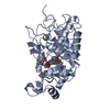 2atjS S: Starting model for refinement |
|---|---|
| Similar structure data |


-Links
 PDBj
PDBj
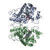
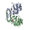
- Assembly
| Deposited unit | 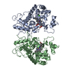
| ||||||||
|---|---|---|---|---|---|---|---|---|---|
| 1 |
| ||||||||
| Unit cell |
| ||||||||
| Noncrystallographic symmetry (NCS) | NCS oper: (Code: given Matrix: (-1, -0.0063, 0.0026), Vector: |
-Components
| #1: Protein | Mass: 34063.363 Da / Num. of mol.: 2 / Mutation: F221M Source method: isolated from a genetically manipulated source Details: CONTAINS HEME, TWO STRUCTURAL CALCIUM IONS, AND BENZHYDROXAMIC ACID BOUND IN ACTIVE SITE Source: (gene. exp.) Armoracia rusticana (horseradish) / Production host:  #2: Chemical | ChemComp-CA /   Mass: 40.078 Da / Num. of mol.: 4 / Source method: obtained synthetically / Formula: Ca Mass: 40.078 Da / Num. of mol.: 4 / Source method: obtained synthetically / Formula: Ca#3: Chemical |   Mass: 616.487 Da / Num. of mol.: 2 / Source method: obtained synthetically / Formula: C34H32FeN4O4 Mass: 616.487 Da / Num. of mol.: 2 / Source method: obtained synthetically / Formula: C34H32FeN4O4#4: Chemical | 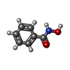  Mass: 137.136 Da / Num. of mol.: 2 / Source method: obtained synthetically / Formula: C7H7NO2 Mass: 137.136 Da / Num. of mol.: 2 / Source method: obtained synthetically / Formula: C7H7NO2#5: Water | ChemComp-HOH / |  Mass: 18.015 Da / Num. of mol.: 364 / Source method: isolated from a natural source / Formula: H2O Mass: 18.015 Da / Num. of mol.: 364 / Source method: isolated from a natural source / Formula: H2OHas protein modification | Y | Nonpolymer details | THE CHARGE IS UNKNOWN SINCE IT IS POSITIONED IN THE ACTIVE SITE, WHICH CHANGES THE NORMAL PKA THE ...THE CHARGE IS UNKNOWN SINCE IT IS POSITIONED | Sequence details | THE N-TERMINAL METHIONINE ORIGINATES FROM THE E. COLI EXPRESSION SYSTEM. RESIDUES 1 - 29 AND 339 - ...THE N-TERMINAL METHIONINE | |
|---|
-Experimental details
-Experiment
| Experiment | Method: X-RAY DIFFRACTION / Number of used crystals: 1 |
|---|
- Sample preparation
| Crystal | Density Matthews: 2.6 Å3/Da / Density % sol: 52.8 % |
|---|---|
| Crystal grow | pH: 6.5 / Details: pH 6.5 |
-Data collection
| Diffraction | Mean temperature: 290 K |
|---|---|
| Diffraction source | Source: ROTATING ANODE / Type: RIGAKU RU200 / Wavelength: 1.5418 |
| Detector | Type: RIGAKU / Detector: IMAGE PLATE / Date: Jun 1, 1997 |
| Radiation | Monochromator: GRAPHITE / Protocol: SINGLE WAVELENGTH / Monochromatic (M) / Laue (L): M / Scattering type: x-ray |
| Radiation wavelength | Wavelength: 1.5418 Å / Relative weight: 1 |
| Reflection | Resolution: 2.2→50 Å / Num. obs: 32731 / % possible obs: 92.2 % / Observed criterion σ(I): -3 / Redundancy: 2.2 % / Biso Wilson estimate: 6.8 Å2 / Rmerge(I) obs: 0.095 / Net I/σ(I): 10 |
| Reflection shell | Resolution: 2.2→2.24 Å / Redundancy: 2 % / Rmerge(I) obs: 0.255 / Mean I/σ(I) obs: 3 / % possible all: 92.1 |
- Processing
| Software |
| ||||||||||||||||||||||||||||||||||||||||||||||||||||||||||||||||||||||||||||||||
|---|---|---|---|---|---|---|---|---|---|---|---|---|---|---|---|---|---|---|---|---|---|---|---|---|---|---|---|---|---|---|---|---|---|---|---|---|---|---|---|---|---|---|---|---|---|---|---|---|---|---|---|---|---|---|---|---|---|---|---|---|---|---|---|---|---|---|---|---|---|---|---|---|---|---|---|---|---|---|---|---|---|
| Refinement | Method to determine structure: MOLECULAR REPLACEMENT Starting model: PDB ENTRY 2ATJ Resolution: 2.2→50 Å / Rfactor Rfree error: 0.004 / Data cutoff high absF: 10000000 / Data cutoff low absF: 0.001 / Isotropic thermal model: RESTRAINED / Cross valid method: THROUGHOUT / σ(F): 0 Details: BULK SOLVENT MODEL USED. THE STRUCTURE WAS REFINED USING STRICT NON- CRYSTALLOGRAPHIC SYMMETRY CONSTRAINTS.
| ||||||||||||||||||||||||||||||||||||||||||||||||||||||||||||||||||||||||||||||||
| Displacement parameters | Biso mean: 15.6 Å2
| ||||||||||||||||||||||||||||||||||||||||||||||||||||||||||||||||||||||||||||||||
| Refine analyze |
| ||||||||||||||||||||||||||||||||||||||||||||||||||||||||||||||||||||||||||||||||
| Refinement step | Cycle: LAST / Resolution: 2.2→50 Å
| ||||||||||||||||||||||||||||||||||||||||||||||||||||||||||||||||||||||||||||||||
| Refine LS restraints |
| ||||||||||||||||||||||||||||||||||||||||||||||||||||||||||||||||||||||||||||||||
| Refine LS restraints NCS | NCS model details: CONSTRAINTS | ||||||||||||||||||||||||||||||||||||||||||||||||||||||||||||||||||||||||||||||||
| LS refinement shell | Resolution: 2.2→2.24 Å / Rfactor Rfree error: 0.02 / Total num. of bins used: 20
| ||||||||||||||||||||||||||||||||||||||||||||||||||||||||||||||||||||||||||||||||
| Xplor file |
|
