 Movie
Movie Controller

[English] 日本語
 Yorodumi
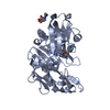
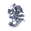
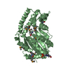
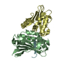
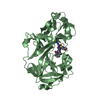
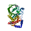
- PDB-2g09: X-ray structure of mouse pyrimidine 5'-nucleotidase type 1, produ... -
+ Open data

- Basic information
| Entry | Database: PDB / ID: 2g09 | ||||||
|---|---|---|---|---|---|---|---|
| Title | X-ray structure of mouse pyrimidine 5'-nucleotidase type 1, product complex | ||||||
 Components Components | Cytosolic 5'-nucleotidase III | ||||||
 Keywords Keywords | HYDROLASE / UniProt Q9D020 / UMPH-1 / Cytosolic 5'-nucleotidase III / Pyrimidine 5'-nucleotidase 1 / P5n-1 / NT5C3 PROTEIN / AAH38029 / BC038029 / MM.158936 / Lead Poisoning / STRUCTURAL GENOMICS FUNCTIONAL FOLLOW-UP STUDY / PROTEIN STRUCTURE INITIATIVE / PSI / CENTER FOR EUKARYOTIC STRUCTURAL GENOMICS / CESG | ||||||
| Function / homology |  Function and homology information Function and homology informationPyrimidine catabolism / dCMP catabolic process / dTMP catabolic process / dUMP catabolic process / UMP catabolic process / 7-methylguanosine nucleotidase / adenosine metabolic process / 5'-nucleotidase / 5'-nucleotidase activity / transferase activity ...Pyrimidine catabolism / dCMP catabolic process / dTMP catabolic process / dUMP catabolic process / UMP catabolic process / 7-methylguanosine nucleotidase / adenosine metabolic process / 5'-nucleotidase / 5'-nucleotidase activity / transferase activity / defense response to virus / nuclear body / nucleotide binding / magnesium ion binding / endoplasmic reticulum / mitochondrion / nucleoplasm / cytosol / cytoplasm Similarity search - Function | ||||||
| Biological species |  | ||||||
| Method |  X-RAY DIFFRACTION / SYNCHROTRON / MOLECULAR REPLACEMENT / Resolution: 2.1 Å X-RAY DIFFRACTION / SYNCHROTRON / MOLECULAR REPLACEMENT / Resolution: 2.1 Å | ||||||
 Authors Authors | Bitto, E. / Bingman, C.A. / Wesenberg, G.E. / Phillips Jr., G.N. / Center for Eukaryotic Structural Genomics (CESG) | ||||||
 Citation Citation | Journal: J.Biol.Chem. / Year: 2006 Title: Structure of pyrimidine 5'-nucleotidase type 1. Insight into mechanism of action and inhibition during lead poisoning. Authors: Bitto, E. / Bingman, C.A. / Wesenberg, G.E. / McCoy, J.G. / Phillips, G.N. | ||||||
| History |
|
- Structure visualization
| Structure viewer | Molecule: MolmilJmol/JSmol |
|---|
- Downloads & links
-Download
| PDBx/mmCIF format | 2g09.cif.gz | 152.6 KB | Display | PDBx/mmCIF format |
|---|---|---|---|---|
| PDB format | pdb2g09.ent.gz | 117.9 KB | Display | PDB format |
| PDBx/mmJSON format | 2g09.json.gz | Tree view | PDBx/mmJSON format | |
| Others |  Other downloads Other downloads |
-Validation report
| Arichive directory | https://data.pdbj.org/pub/pdb/validation_reports/g0/2g09ftp://data.pdbj.org/pub/pdb/validation_reports/g0/2g09 | HTTPS FTP |
|---|
-Related structure data
| Related structure data |  2bduSC  2g06C  2g07C 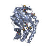 2g08C  2g0aC S: Starting model for refinement C: citing same article ( |
|---|---|
| Similar structure data | |
| Other databases |


-Links
 PDBj
PDBj
- Assembly
| Deposited unit | 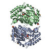
| ||||||||||||||||||
|---|---|---|---|---|---|---|---|---|---|---|---|---|---|---|---|---|---|---|---|
| 1 | 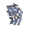
| ||||||||||||||||||
| 2 | 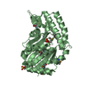
| ||||||||||||||||||
| Unit cell |
| ||||||||||||||||||
| Noncrystallographic symmetry (NCS) | NCS domain:
NCS domain segments: Component-ID: 1 / Ens-ID: 1 / Beg auth comp-ID: ALA / Beg label comp-ID: ALA / End auth comp-ID: LEU / End label comp-ID: LEU / Refine code: 2 / Auth seq-ID: 7 - 297 / Label seq-ID: 7 - 297
| ||||||||||||||||||
| Details | The biological unit is a monomer. There are 2 biological units in the asymmetric unit (chains A & B). |
-Components
| #1: Protein | Mass: 34163.926 Da / Num. of mol.: 2 Source method: isolated from a genetically manipulated source Source: (gene. exp.)  #2: Chemical |   Mass: 24.305 Da / Num. of mol.: 2 / Source method: obtained synthetically / Formula: Mg Mass: 24.305 Da / Num. of mol.: 2 / Source method: obtained synthetically / Formula: Mg#3: Chemical |   Mass: 94.971 Da / Num. of mol.: 2 / Source method: obtained synthetically / Formula: PO4 Mass: 94.971 Da / Num. of mol.: 2 / Source method: obtained synthetically / Formula: PO4#4: Chemical | 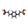  Mass: 302.368 Da / Num. of mol.: 2 / Source method: obtained synthetically / Formula: C8H18N2O6S2 / Comment: pH buffer*YM Mass: 302.368 Da / Num. of mol.: 2 / Source method: obtained synthetically / Formula: C8H18N2O6S2 / Comment: pH buffer*YM#5: Water | ChemComp-HOH / |  Mass: 18.015 Da / Num. of mol.: 848 / Source method: isolated from a natural source / Formula: H2O Mass: 18.015 Da / Num. of mol.: 848 / Source method: isolated from a natural source / Formula: H2OHas protein modification | Y | |
|---|
-Experimental details
-Experiment
| Experiment | Method: X-RAY DIFFRACTION / Number of used crystals: 1 |
|---|
- Sample preparation
| Crystal | Density Matthews: 2.96 Å3/Da / Density % sol: 58.51 % |
|---|---|
| Crystal grow | Temperature: 277 K / Method: vapor diffusion, hanging drop / pH: 6 Details: PROTEIN SOLUTION (10 MG/ML PROTEIN, 0.005 M BIS TRIS, 0.050 M SODIUM CHLORIDE, 0.003 M SODIUM AZIDE, 0.0003 M TCEP, PH 6.0) MIXED IN A 1:1 RATIO WITH THE WELL SOLUTION (20-25% PEG 8K, 0.10 M ...Details: PROTEIN SOLUTION (10 MG/ML PROTEIN, 0.005 M BIS TRIS, 0.050 M SODIUM CHLORIDE, 0.003 M SODIUM AZIDE, 0.0003 M TCEP, PH 6.0) MIXED IN A 1:1 RATIO WITH THE WELL SOLUTION (20-25% PEG 8K, 0.10 M PIPES PH 6.5) CRYSTALS SOAKED FOR 60 MINUTES IN WELL SOLUTION WITH 0.005 M magnesium chloride, 0.005 M UMP, temperature 277K, VAPOR DIFFUSION, HANGING DROP, pH 6.00 |
-Data collection
| Diffraction | Mean temperature: 100 K |
|---|---|
| Diffraction source | Source: SYNCHROTRON / Site: APS  / Beamline: 8-BM / Wavelength: 0.97913 / Beamline: 8-BM / Wavelength: 0.97913 |
| Detector | Type: ADSC QUANTUM 315 / Detector: CCD / Date: Nov 30, 2005 |
| Radiation | Monochromator: SI 111 / Protocol: SINGLE WAVELENGTH / Monochromatic (M) / Laue (L): M / Scattering type: x-ray |
| Radiation wavelength | Wavelength: 0.97913 Å / Relative weight: 1 |
| Reflection | Resolution: 2.1→50 Å / Num. obs: 45396 / % possible obs: 98.9 % / Redundancy: 3.8 % / Rmerge(I) obs: 0.071 / Net I/σ(I): 13.11 |
| Reflection shell | Resolution: 2.1→2.15 Å / Redundancy: 3.2 % / Rmerge(I) obs: 0.342 / Mean I/σ(I) obs: 3.42 / % possible all: 97.6 |
- Processing
| Software |
| ||||||||||||||||||||||||||||||||||||||||||||||||||||||||||||||||||||||||||||||||||||||||||||||||||||||||||||||||||||||||||||||||||||||||||||||||||||||||||||||||||||||||||
|---|---|---|---|---|---|---|---|---|---|---|---|---|---|---|---|---|---|---|---|---|---|---|---|---|---|---|---|---|---|---|---|---|---|---|---|---|---|---|---|---|---|---|---|---|---|---|---|---|---|---|---|---|---|---|---|---|---|---|---|---|---|---|---|---|---|---|---|---|---|---|---|---|---|---|---|---|---|---|---|---|---|---|---|---|---|---|---|---|---|---|---|---|---|---|---|---|---|---|---|---|---|---|---|---|---|---|---|---|---|---|---|---|---|---|---|---|---|---|---|---|---|---|---|---|---|---|---|---|---|---|---|---|---|---|---|---|---|---|---|---|---|---|---|---|---|---|---|---|---|---|---|---|---|---|---|---|---|---|---|---|---|---|---|---|---|---|---|---|---|---|---|
| Refinement | Method to determine structure: MOLECULAR REPLACEMENT Starting model: PDB ENTRY 2BDU Resolution: 2.1→32.19 Å / Cor.coef. Fo:Fc: 0.97 / Cor.coef. Fo:Fc free: 0.953 / SU B: 6.969 / SU ML: 0.104 / TLS residual ADP flag: LIKELY RESIDUAL / Cross valid method: THROUGHOUT / ESU R: 0.166 / ESU R Free: 0.152 / Details: HYDROGENS HAVE BEEN ADDED IN THE RIDING POSITIONS
| ||||||||||||||||||||||||||||||||||||||||||||||||||||||||||||||||||||||||||||||||||||||||||||||||||||||||||||||||||||||||||||||||||||||||||||||||||||||||||||||||||||||||||
| Solvent computation | Ion probe radii: 0.8 Å / Shrinkage radii: 0.8 Å / VDW probe radii: 1.2 Å / Solvent model: MASK BULK SOLVENT | ||||||||||||||||||||||||||||||||||||||||||||||||||||||||||||||||||||||||||||||||||||||||||||||||||||||||||||||||||||||||||||||||||||||||||||||||||||||||||||||||||||||||||
| Displacement parameters | Biso mean: 38.09 Å2
| ||||||||||||||||||||||||||||||||||||||||||||||||||||||||||||||||||||||||||||||||||||||||||||||||||||||||||||||||||||||||||||||||||||||||||||||||||||||||||||||||||||||||||
| Refinement step | Cycle: LAST / Resolution: 2.1→32.19 Å
| ||||||||||||||||||||||||||||||||||||||||||||||||||||||||||||||||||||||||||||||||||||||||||||||||||||||||||||||||||||||||||||||||||||||||||||||||||||||||||||||||||||||||||
| Refine LS restraints |
| ||||||||||||||||||||||||||||||||||||||||||||||||||||||||||||||||||||||||||||||||||||||||||||||||||||||||||||||||||||||||||||||||||||||||||||||||||||||||||||||||||||||||||
| Refine LS restraints NCS | Dom-ID: 1 / Auth asym-ID: A / Ens-ID: 1 / Refine-ID: X-RAY DIFFRACTION
| ||||||||||||||||||||||||||||||||||||||||||||||||||||||||||||||||||||||||||||||||||||||||||||||||||||||||||||||||||||||||||||||||||||||||||||||||||||||||||||||||||||||||||
| LS refinement shell | Resolution: 2.1→2.15 Å / Total num. of bins used: 20
| ||||||||||||||||||||||||||||||||||||||||||||||||||||||||||||||||||||||||||||||||||||||||||||||||||||||||||||||||||||||||||||||||||||||||||||||||||||||||||||||||||||||||||
| Refinement TLS params. | Method: refined / Refine-ID: X-RAY DIFFRACTION
| ||||||||||||||||||||||||||||||||||||||||||||||||||||||||||||||||||||||||||||||||||||||||||||||||||||||||||||||||||||||||||||||||||||||||||||||||||||||||||||||||||||||||||
| Refinement TLS group |
|
