 Movie
Movie Controller

[English] 日本語
 Yorodumi
- PDB-6t9m: Crystal structure of the Chitinase Domain of the Spore Coat Prote... -
+ Open data

- Basic information
| Entry | Database: PDB / ID: 6t9m | ||||||
|---|---|---|---|---|---|---|---|
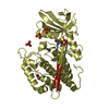
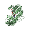
| Title | Crystal structure of the Chitinase Domain of the Spore Coat Protein CotE from Clostridium difficile | ||||||
 Components Components |
| ||||||
 Keywords Keywords | STRUCTURAL PROTEIN / Spore Coat / Chitinase / Colonisation Factor | ||||||
| Function / homology |  Function and homology information Function and homology informationthioredoxin-dependent peroxiredoxin activity / thioredoxin-dependent peroxiredoxin / endochitinase activity / chitin catabolic process / chitin binding / carbohydrate metabolic process / extracellular region / cytoplasm Similarity search - Function | ||||||
| Biological species |  Peptoclostridium difficile (bacteria) Peptoclostridium difficile (bacteria) | ||||||
| Method |  X-RAY DIFFRACTION / SYNCHROTRON / MOLECULAR REPLACEMENT / Resolution: 1.3 Å X-RAY DIFFRACTION / SYNCHROTRON / MOLECULAR REPLACEMENT / Resolution: 1.3 Å | ||||||
 Authors Authors | Whittingham, J.L. / Dodson, E.J. / Wilkinson, A.J. | ||||||
| Funding support |  United Kingdom, 1items United Kingdom, 1items
| ||||||
 Citation Citation | Journal: Acta Crystallogr.,Sect.F / Year: 2020 Title: Crystal structures of the GH18 domain of the bifunctional peroxiredoxin-chitinase CotE from Clostridium difficile. Authors: Whittingham, J.L. / Hanai, S. / Brannigan, J.A. / Ferreira, W.T. / Dodson, E.J. / Turkenburg, J.P. / Cartwright, J. / Cutting, S.M. / Wilkinson, A.J. #1: Journal: J. Infect. Dis. / Year: 2017 Title: The Spore Coat Protein CotE Facilitates Host Colonization by Clostridium difficile. Authors: Hong, H.A. / Ferreira, W.T. / Hosseini, S. / Anwar, S. / Hitri, K. / Wilkinson, A.J. / Vahjen, W. / Zentek, J. / Soloviev, M. / Cutting, S.M. | ||||||
| History |
|
- Structure visualization
| Structure viewer | Molecule: MolmilJmol/JSmol |
|---|
- Downloads & links
-Download
| PDBx/mmCIF format | 6t9m.cif.gz | 260.5 KB | Display | PDBx/mmCIF format |
|---|---|---|---|---|
| PDB format | pdb6t9m.ent.gz | Display | PDB format | |
| PDBx/mmJSON format | 6t9m.json.gz | Tree view | PDBx/mmJSON format | |
| Others |  Other downloads Other downloads |
-Validation report
| Arichive directory | https://data.pdbj.org/pub/pdb/validation_reports/t9/6t9mftp://data.pdbj.org/pub/pdb/validation_reports/t9/6t9m | HTTPS FTP |
|---|
-Related structure data


-Links
 PDBj
PDBj- Assembly
| Deposited unit | 
| ||||||||
|---|---|---|---|---|---|---|---|---|---|
| 1 | 
| ||||||||
| 2 | 
| ||||||||
| Unit cell |
|
-Components
| #1: Protein | Mass: 42924.008 Da / Num. of mol.: 1 Source method: isolated from a genetically manipulated source Details: This is the C-terminal chitinase domain of a bifunctional protein found in the spores of Clostridium difficile. It has an HRV 3C cleavable H6 tag fused to its N-terminus Source: (gene. exp.) Peptoclostridium difficile (strain 630) (bacteria)Strain: 630 / Gene: cotE, CD630_14330 / Plasmid: pETYSBLLIC3C Details (production host): T7 promoter regulated by lac repressor/operator Production host: | ||||||||
|---|---|---|---|---|---|---|---|---|---|
| #2: Protein/peptide | Mass: 503.635 Da / Num. of mol.: 1 / Source method: obtained synthetically / Details: peptide defined by electron density in active site / Source: (synth.) | ||||||||
| #3: Chemical |   Mass: 238.278 Da / Num. of mol.: 2 / Source method: isolated from a natural source / Formula: C10H22O6 / Comment: precipitant*YM Mass: 238.278 Da / Num. of mol.: 2 / Source method: isolated from a natural source / Formula: C10H22O6 / Comment: precipitant*YM#4: Chemical |   Mass: 106.120 Da / Num. of mol.: 3 / Source method: obtained synthetically / Formula: C4H10O3 Mass: 106.120 Da / Num. of mol.: 3 / Source method: obtained synthetically / Formula: C4H10O3#5: Water | ChemComp-HOH / |  Mass: 18.015 Da / Num. of mol.: 490 / Source method: isolated from a natural source / Formula: H2O Mass: 18.015 Da / Num. of mol.: 490 / Source method: isolated from a natural source / Formula: H2OHas ligand of interest | N | Has protein modification | Y | |
-Experimental details
-Experiment
| Experiment | Method: X-RAY DIFFRACTION / Number of used crystals: 1 |
|---|
- Sample preparation
| Crystal | Density Matthews: 2.29 Å3/Da / Density % sol: 46.21 % |
|---|---|
| Crystal grow | Temperature: 291 K / Method: vapor diffusion, hanging drop / pH: 8 Details: Well solution: 200 mM ammonium phosphate, 22.5% PEG 3350 Protein solution: 20 mM Tris-HCl, pH 8.0, 150 mM NaCl Drop: 3 microlitre (1 microlitre protein plus 2 microlitre reservoir solution) |
-Data collection
| Diffraction | Mean temperature: 100 K / Serial crystal experiment: N |
|---|---|
| Diffraction source | Source: SYNCHROTRON / Site: Diamond / Beamline: I02 / Wavelength: 0.9795 Å |
| Detector | Type: DECTRIS PILATUS3 2M / Detector: PIXEL / Date: Feb 1, 2016 |
| Radiation | Protocol: SINGLE WAVELENGTH / Monochromatic (M) / Laue (L): M / Scattering type: x-ray |
| Radiation wavelength | Wavelength: 0.9795 Å / Relative weight: 1 |
| Reflection | Resolution: 1.3→47.21 Å / Num. obs: 94650 / % possible obs: 97.9 % / Redundancy: 4.6 % / CC1/2: 0.997 / Rrim(I) all: 0.058 / Net I/σ(I): 18 |
| Reflection shell | Resolution: 1.3→1.32 Å / Redundancy: 4.7 % / Mean I/σ(I) obs: 6.5 / Num. unique obs: 4564 / CC1/2: 0.954 / Rrim(I) all: 0.31 / % possible all: 96.1 |
- Processing
| Software |
| ||||||||||||||||||||||||||||||||||||||||||||||||||||||||||||||||||||||||||||||||||||||||||||||||||||||||||||||||||||||||||||||||||||||||||||||||||||||||||||||||
|---|---|---|---|---|---|---|---|---|---|---|---|---|---|---|---|---|---|---|---|---|---|---|---|---|---|---|---|---|---|---|---|---|---|---|---|---|---|---|---|---|---|---|---|---|---|---|---|---|---|---|---|---|---|---|---|---|---|---|---|---|---|---|---|---|---|---|---|---|---|---|---|---|---|---|---|---|---|---|---|---|---|---|---|---|---|---|---|---|---|---|---|---|---|---|---|---|---|---|---|---|---|---|---|---|---|---|---|---|---|---|---|---|---|---|---|---|---|---|---|---|---|---|---|---|---|---|---|---|---|---|---|---|---|---|---|---|---|---|---|---|---|---|---|---|---|---|---|---|---|---|---|---|---|---|---|---|---|---|---|---|---|
| Refinement | Method to determine structure: MOLECULAR REPLACEMENT Starting model: xxx Resolution: 1.3→45.09 Å / Cor.coef. Fo:Fc: 0.983 / Cor.coef. Fo:Fc free: 0.975 / WRfactor Rfree: 0.137 / WRfactor Rwork: 0.107 / Average fsc free: 0.9794 / Average fsc work: 0.9835 / Cross valid method: FREE R-VALUE / ESU R: 0.034 / ESU R Free: 0.035 Details: Hydrogens have been added in their riding positions
| ||||||||||||||||||||||||||||||||||||||||||||||||||||||||||||||||||||||||||||||||||||||||||||||||||||||||||||||||||||||||||||||||||||||||||||||||||||||||||||||||
| Solvent computation | Ion probe radii: 0.8 Å / Shrinkage radii: 0.8 Å / VDW probe radii: 1.2 Å / Solvent model: MASK BULK SOLVENT | ||||||||||||||||||||||||||||||||||||||||||||||||||||||||||||||||||||||||||||||||||||||||||||||||||||||||||||||||||||||||||||||||||||||||||||||||||||||||||||||||
| Displacement parameters | Biso mean: 14.194 Å2
| ||||||||||||||||||||||||||||||||||||||||||||||||||||||||||||||||||||||||||||||||||||||||||||||||||||||||||||||||||||||||||||||||||||||||||||||||||||||||||||||||
| Refinement step | Cycle: LAST / Resolution: 1.3→45.09 Å
| ||||||||||||||||||||||||||||||||||||||||||||||||||||||||||||||||||||||||||||||||||||||||||||||||||||||||||||||||||||||||||||||||||||||||||||||||||||||||||||||||
| Refine LS restraints |
| ||||||||||||||||||||||||||||||||||||||||||||||||||||||||||||||||||||||||||||||||||||||||||||||||||||||||||||||||||||||||||||||||||||||||||||||||||||||||||||||||
| LS refinement shell |
|
