[English] 日本語
Yorodumi
- PDB-6vzh: Structure of Human Vaccinia-related Kinase 1 (VRK1) Bound to LDSM311 -

+
Open data


ID or keywords:

Loading...

-
Basic information

Entry
Database: PDB / ID: 6vzh
TitleStructure of Human Vaccinia-related Kinase 1 (VRK1) Bound to LDSM311
ComponentsSerine/threonine-protein kinase VRK1
KeywordsTRANSFERASE/TRANSFERASE INHIBITOR / protein kinase / inhibitor / Structural Genomics / Structural Genomics Consortium / SGC / TRANSFERASE-TRANSFERASE INHIBITOR / TRANSFERASE-TRANSFERASE INHIBITOR complex
Function / homology
Function and homology information


Golgi disassembly / histone H3T3 kinase activity / Nuclear Envelope Breakdown / positive regulation of protein localization to chromatin / mitotic nuclear membrane disassembly / Golgi stack / Initiation of Nuclear Envelope (NE) Reformation / histone H3S10 kinase activity / kinase activity / histone binding ...Golgi disassembly / histone H3T3 kinase activity / Nuclear Envelope Breakdown / positive regulation of protein localization to chromatin / mitotic nuclear membrane disassembly / Golgi stack / Initiation of Nuclear Envelope (NE) Reformation / histone H3S10 kinase activity / kinase activity / histone binding / protein autophosphorylation / non-specific serine/threonine protein kinase / protein kinase activity / cell division / protein phosphorylation / protein serine kinase activity / protein serine/threonine kinase activity / DNA damage response / nucleolus / protein kinase binding / signal transduction / nucleoplasm / ATP binding / nucleus / cytosol
Similarity search - Function
Transferase(Phosphotransferase) domain 1 / Transferase(Phosphotransferase); domain 1 / Serine/threonine-protein kinase, active site / Serine/Threonine protein kinases active-site signature. / Protein kinase domain / Serine/Threonine protein kinases, catalytic domain / Protein kinase, ATP binding site / Protein kinases ATP-binding region signature. / Protein kinase domain profile. / Protein kinase domain ...Transferase(Phosphotransferase) domain 1 / Transferase(Phosphotransferase); domain 1 / Serine/threonine-protein kinase, active site / Serine/Threonine protein kinases active-site signature. / Protein kinase domain / Serine/Threonine protein kinases, catalytic domain / Protein kinase, ATP binding site / Protein kinases ATP-binding region signature. / Protein kinase domain profile. / Protein kinase domain / Protein kinase-like domain superfamily / Orthogonal Bundle / Mainly Alpha
Similarity search - Domain/homology
Chem-RYA / Serine/threonine-protein kinase VRK1
Similarity search - Component
Biological speciesHomo sapiens (human)
MethodX-RAY DIFFRACTION / SYNCHROTRON / MOLECULAR REPLACEMENT / molecular replacement / Resolution: 2.55 Å
Authorsdos Reis, C.V. / Dutra, L.A. / Gama, F. / Ferreira, M. / Mascarello, A. / Azevedo, H. / Guimaraes, C. / Massirer, K.B. / Arruda, P. / Edwards, A.M. ...dos Reis, C.V. / Dutra, L.A. / Gama, F. / Ferreira, M. / Mascarello, A. / Azevedo, H. / Guimaraes, C. / Massirer, K.B. / Arruda, P. / Edwards, A.M. / Counago, R.M. / Structural Genomics Consortium (SGC)
Funding support Brazil, 3items
OrganizationGrant numberCountry
Sao Paulo Research Foundation (FAPESP)13/50724-5 Brazil
Brazilian National Council for Scientific and Technological Development (CNPq)465651/2014-3 Brazil
Sao Paulo Research Foundation (FAPESP)2014/50897-0 Brazil
CitationJournal: To be Published
Title: Structure of Human Vaccinia-related Kinase 1 (VRK1) Bound to LDSM311
Authors: dos Reis, C.V. / Dutra, L.A. / Gama, F. / Ferreira, M. / Mascarello, A. / Azevedo, H. / Guimaraes, C.R.W. / Massirer, K.B. / Arruda, P. / Edwards, A.M. / Counago, R.M.
History
DepositionFeb 28, 2020Deposition site: RCSB / Processing site: RCSB
Revision 1.0Mar 11, 2020Provider: repository / Type: Initial release
Revision 1.1Oct 11, 2023Group: Data collection / Database references / Refinement description
Category: chem_comp_atom / chem_comp_bond ...chem_comp_atom / chem_comp_bond / database_2 / pdbx_initial_refinement_model
Item: _database_2.pdbx_DOI / _database_2.pdbx_database_accession

-
Structure visualization

Structure viewerMolecule:
MolmilJmol/JSmol

Downloads & links

-
Assembly

Deposited unit
A: Serine/threonine-protein kinase VRK1
B: Serine/threonine-protein kinase VRK1
C: Serine/threonine-protein kinase VRK1
D: Serine/threonine-protein kinase VRK1
hetero molecules


Theoretical massNumber of molelcules
Total (without water)166,19816
Polymers164,5534
Non-polymers1,64512
Water57632
1
A: Serine/threonine-protein kinase VRK1
hetero molecules


Theoretical massNumber of molelcules
Total (without water)41,3303
Polymers41,1381
Non-polymers1922
Water181
TypeNameSymmetry operationNumber
identity operation1_555x,y,z1
2
B: Serine/threonine-protein kinase VRK1
hetero molecules


Theoretical massNumber of molelcules
Total (without water)41,7865
Polymers41,1381
Non-polymers6484
Water181
TypeNameSymmetry operationNumber
identity operation1_555x,y,z1
3
C: Serine/threonine-protein kinase VRK1
hetero molecules


Theoretical massNumber of molelcules
Total (without water)41,3303
Polymers41,1381
Non-polymers1922
Water181
TypeNameSymmetry operationNumber
identity operation1_555x,y,z1
4
D: Serine/threonine-protein kinase VRK1
hetero molecules


Theoretical massNumber of molelcules
Total (without water)41,7525
Polymers41,1381
Non-polymers6144
Water181
TypeNameSymmetry operationNumber
identity operation1_555x,y,z1
Unit cell
Length a, b, c (Å)92.158, 95.641, 192.593
Angle α, β, γ (deg.)90.000, 90.000, 90.000
Int Tables number19
Space group name H-MP212121

-
Components

#1: Protein
Serine/threonine-protein kinase VRK1 / Vaccinia-related kinase 1


Mass: 41138.125 Da / Num. of mol.: 4
Mutation: K34A,K35A,E36A,E212A,K214A,E215A,E292A,K293A,K295A,K359A,K360A
Source method: isolated from a genetically manipulated source
Source: (gene. exp.) Homo sapiens (human) / Gene: VRK1 / Production host: Escherichia coli BL21(DE3) (bacteria) / Strain (production host): R3
References: UniProt: Q99986, non-specific serine/threonine protein kinase
#2: Chemical
ChemComp-SO4 / SULFATE ION / Sulfate


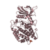
Mass: 96.063 Da / Num. of mol.: 9 / Source method: obtained synthetically / Formula: SO4
#3: Chemical ChemComp-RYA / (7~{R})-2-[[3,5-bis(fluoranyl)-4-oxidanyl-phenyl]amino]-5,7-dimethyl-8-prop-2-ynyl-7~{H}-pteridin-6-one


Mass: 359.330 Da / Num. of mol.: 2 / Source method: obtained synthetically / Formula: C17H15F2N5O2 / Feature type: SUBJECT OF INVESTIGATION
#4: Chemical ChemComp-EDO / 1,2-ETHANEDIOL / ETHYLENE GLYCOL / Ethylene glycol


Mass: 62.068 Da / Num. of mol.: 1 / Source method: obtained synthetically / Formula: C2H6O2
#5: Water ChemComp-HOH / water / Water


Mass: 18.015 Da / Num. of mol.: 32 / Source method: isolated from a natural source / Formula: H2O
Has ligand of interestY

-
Experimental details

-
Experiment

ExperimentMethod: X-RAY DIFFRACTION / Number of used crystals: 1

-
Sample preparation

CrystalDensity Matthews: 2.58 Å3/Da / Density % sol: 52.31 %
Crystal growTemperature: 293 K / Method: vapor diffusion, sitting drop / pH: 6.5
Details: 27.5% PEG3350, 300 mM lithium sulfate, 0.1 M sodium tartrate, 0.1 M Bis-Tris, 0.1 M glycylglycine, cryoprotectant: 30% glycerol

-
Data collection

DiffractionMean temperature: 100 K / Serial crystal experiment: N
Diffraction sourceSource: SYNCHROTRON / Site: APS / Beamline: 24-ID-C / Wavelength: 0.97918 Å
DetectorType: DECTRIS PILATUS 6M-F / Detector: PIXEL / Date: Dec 5, 2019
RadiationMonochromator: cryo-cooled double crystal Si(111) / Protocol: SINGLE WAVELENGTH / Monochromatic (M) / Laue (L): M / Scattering type: x-ray
Radiation wavelengthWavelength: 0.97918 Å / Relative weight: 1
ReflectionResolution: 2.55→96.3 Å / Num. obs: 56320 / % possible obs: 99.9 % / Redundancy: 6.7 % / CC1/2: 0.997 / Rmerge(I) obs: 0.131 / Rpim(I) all: 0.055 / Rrim(I) all: 0.142 / Net I/σ(I): 8.7 / Num. measured all: 376419 / Scaling rejects: 6
Reflection shell

Diffraction-ID: 1

Resolution (Å)Redundancy (%)Rmerge(I) obsNum. measured allNum. unique obsCC1/2Rpim(I) allRrim(I) allNet I/σ(I) obs% possible all
2.55-2.626.91.5593156545690.6760.6371.6861.199.9
10.82-96.35.90.04750328580.9980.0210.05225.599.8

-
Phasing

PhasingMethod: molecular replacement

-
Processing

Software
NameVersionClassificationNB
XDSdata reduction
Aimless0.7.4data scaling
PHASERphasing
REFMAC5.8.0238refinement
PDB_EXTRACT3.25data extraction
RefinementMethod to determine structure: MOLECULAR REPLACEMENT
Starting model: PDB entry 6BRU
Resolution: 2.55→20 Å / Cor.coef. Fo:Fc: 0.951 / Cor.coef. Fo:Fc free: 0.931 / SU B: 30.444 / SU ML: 0.28 / SU R Cruickshank DPI: 0.4584 / Cross valid method: THROUGHOUT / σ(F): 0 / ESU R: 0.457 / ESU R Free: 0.278
Details: HYDROGENS HAVE BEEN ADDED IN THE RIDING POSITIONS U VALUES : WITH TLS ADDED
RfactorNum. reflection% reflectionSelection details
Rfree0.2514 2757 4.9 %RANDOM
Rwork0.2157 ---
obs0.2174 53338 99.63 %-
Solvent computationIon probe radii: 0.8 Å / Shrinkage radii: 0.8 Å / VDW probe radii: 1.2 Å
Displacement parametersBiso max: 143.64 Å2 / Biso mean: 63.234 Å2 / Biso min: 43.02 Å2
Baniso -1Baniso -2Baniso -3
1--0.54 Å20 Å20 Å2
2--2.27 Å2-0 Å2
3----1.73 Å2
Refinement stepCycle: final / Resolution: 2.55→20 Å
ProteinNucleic acidLigandSolventTotal
Num. atoms9562 0 101 32 9695
Biso mean--97.49 58.71 -
Num. residues----1244
Refine LS restraints
Refine-IDTypeDev idealDev ideal targetNumber
X-RAY DIFFRACTIONr_bond_refined_d0.0070.0139886
X-RAY DIFFRACTIONr_bond_other_d0.0020.0178843
X-RAY DIFFRACTIONr_angle_refined_deg1.4891.64113461
X-RAY DIFFRACTIONr_angle_other_deg1.251.57720336
X-RAY DIFFRACTIONr_dihedral_angle_1_deg6.86551232
X-RAY DIFFRACTIONr_dihedral_angle_2_deg28.07921.883494
X-RAY DIFFRACTIONr_dihedral_angle_3_deg16.016151492
X-RAY DIFFRACTIONr_dihedral_angle_4_deg17.9561559
X-RAY DIFFRACTIONr_chiral_restr0.0650.21275
X-RAY DIFFRACTIONr_gen_planes_refined0.0060.0211191
X-RAY DIFFRACTIONr_gen_planes_other0.0010.022126
LS refinement shellResolution: 2.55→2.615 Å / Rfactor Rfree error: 0 / Total num. of bins used: 20
RfactorNum. reflection% reflection
Rfree0.356 231 -
Rwork0.35 3804 -
all-4035 -
obs--99.9 %
Refinement TLS params.

Method: refined / Refine-ID: X-RAY DIFFRACTION

IDL112)L122)L132)L222)L232)L332)S11 (Å °)S12 (Å °)S13 (Å °)S21 (Å °)S22 (Å °)S23 (Å °)S31 (Å °)S32 (Å °)S33 (Å °)T112)T122)T132)T222)T232)T332)Origin x (Å)Origin y (Å)Origin z (Å)
11.4902-0.5493-1.44831.67591.22533.28590.03960.07740.11660.1294-0.0532-0.077-0.0998-0.11710.01350.3611-0.0114-0.00220.35480.00550.023218.45342.439220.9211
22.7455-0.20990.31212.2253-0.39021.8604-0.0390.0319-0.1376-0.01370.08120.3913-0.031-0.418-0.04220.46540.00970.02390.4573-0.00860.0751-15.13082.497552.7564
31.1593-0.00630.92211.7739-0.67553.60430.0163-0.0365-0.06380.1524-0.05050.2320.2128-0.37090.03420.2943-0.08280.03940.4279-0.03730.0453-25.8355-45.007727.7049
42.6237-0.5114-0.82452.37040.80742.9654-0.0143-0.12470.53060.12460.1489-0.2452-0.13010.1107-0.13460.29740.0047-0.03580.4287-0.01970.1201-26.689-9.798-2.6565
Refinement TLS group
IDRefine-IDRefine TLS-IDAuth asym-IDAuth seq-ID
1X-RAY DIFFRACTION1A20 - 402
2X-RAY DIFFRACTION2B23 - 501
3X-RAY DIFFRACTION3C21 - 402
4X-RAY DIFFRACTION4D22 - 501

+
About Yorodumi

-
News

-
Feb 9, 2022. New format data for meta-information of EMDB entries

New format data for meta-information of EMDB entries

  • Version 3 of the EMDB header file is now the official format.
  • The previous official version 1.9 will be removed from the archive.

Related info.:EMDB header

External links:wwPDB to switch to version 3 of the EMDB data model

-
Aug 12, 2020. Covid-19 info

Covid-19 info

URL: https://pdbj.org/emnavi/covid19.php

New page: Covid-19 featured information page in EM Navigator.

Related info.:Covid-19 info / Mar 5, 2020. Novel coronavirus structure data

+
Mar 5, 2020. Novel coronavirus structure data

Novel coronavirus structure data

Related info.:Yorodumi Speices / Aug 12, 2020. Covid-19 info

External links:COVID-19 featured content - PDBj / Molecule of the Month (242):Coronavirus Proteases

+
Jan 31, 2019. EMDB accession codes are about to change! (news from PDBe EMDB page)

EMDB accession codes are about to change! (news from PDBe EMDB page)

  • The allocation of 4 digits for EMDB accession codes will soon come to an end. Whilst these codes will remain in use, new EMDB accession codes will include an additional digit and will expand incrementally as the available range of codes is exhausted. The current 4-digit format prefixed with “EMD-” (i.e. EMD-XXXX) will advance to a 5-digit format (i.e. EMD-XXXXX), and so on. It is currently estimated that the 4-digit codes will be depleted around Spring 2019, at which point the 5-digit format will come into force.
  • The EM Navigator/Yorodumi systems omit the EMD- prefix.

Related info.:Q: What is EMD? / ID/Accession-code notation in Yorodumi/EM Navigator

External links:EMDB Accession Codes are Changing Soon! / Contact to PDBj

+
Jul 12, 2017. Major update of PDB

Major update of PDB

  • wwPDB released updated PDB data conforming to the new PDBx/mmCIF dictionary.
  • This is a major update changing the version number from 4 to 5, and with Remediation, in which all the entries are updated.
  • In this update, many items about electron microscopy experimental information are reorganized (e.g. em_software).
  • Now, EM Navigator and Yorodumi are based on the updated data.

External links:wwPDB Remediation / Enriched Model Files Conforming to OneDep Data Standards Now Available in the PDB FTP Archive

-
Yorodumi

Thousand views of thousand structures

  • Yorodumi is a browser for structure data from EMDB, PDB, SASBDB, etc.
  • This page is also the successor to EM Navigator detail page, and also detail information page/front-end page for Omokage search.
  • The word "yorodu" (or yorozu) is an old Japanese word meaning "ten thousand". "mi" (miru) is to see.

Related info.:EMDB / PDB / SASBDB / Comparison of 3 databanks / Yorodumi Search / Aug 31, 2016. New EM Navigator & Yorodumi / Yorodumi Papers / Jmol/JSmol / Function and homology information / Changes in new EM Navigator and Yorodumi

Read more