 Movie
Movie Controller

[English] 日本語
 Yorodumi
- PDB-1jqa: Bacillus stearothermophilus glycerol dehydrogenase complex with g... -
+ Open data

- Basic information
| Entry | Database: PDB / ID: 1jqa | ||||||
|---|---|---|---|---|---|---|---|
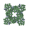
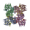
| Title | Bacillus stearothermophilus glycerol dehydrogenase complex with glycerol | ||||||
 Components Components | Glycerol Dehydrogenase | ||||||
 Keywords Keywords | OXIDOREDUCTASE / NAD / Glycerol metabolism | ||||||
| Function / homology |  Function and homology information Function and homology informationanaerobic glycerol catabolic process / glycerol dehydrogenase / glycerol dehydrogenase (NAD+) activity / metal ion binding / cytosol Similarity search - Function | ||||||
| Biological species |   Geobacillus stearothermophilus (bacteria) Geobacillus stearothermophilus (bacteria) | ||||||
| Method |  X-RAY DIFFRACTION / SYNCHROTRON / MOLECULAR REPLACEMENT / Resolution: 2.05 Å X-RAY DIFFRACTION / SYNCHROTRON / MOLECULAR REPLACEMENT / Resolution: 2.05 Å | ||||||
 Authors Authors | Ruzheinikov, S.N. / Burke, J. / Sedelnikova, S. / Baker, P.J. / Taylor, R. / Bullough, P.A. / Muir, N.M. / Gore, M.G. / Rice, D.W. | ||||||
 Citation Citation | Journal: Structure / Year: 2001 Title: Glycerol dehydrogenase. structure, specificity, and mechanism of a family III polyol dehydrogenase. Authors: Ruzheinikov, S.N. / Burke, J. / Sedelnikova, S. / Baker, P.J. / Taylor, R. / Bullough, P.A. / Muir, N.M. / Gore, M.G. / Rice, D.W. #1: Journal: Acta Crystallogr.,Sect.D / Year: 2001Title: Purification, crystallization and quaternary structure analysis of a glycerol dehydrogenase S305C mutant from Bacillus stearothemophilus. Authors: Burke, J. / Ruzheinikov, S.N. / Sedelnikova, S. / Baker, P.J. / Holmes, D. / Muir, N.M. / Gore, M.G. / Rice, D.W. | ||||||
| History |
|
- Structure visualization
| Structure viewer | Molecule: MolmilJmol/JSmol |
|---|
- Downloads & links
-Download
| PDBx/mmCIF format | 1jqa.cif.gz | 86 KB | Display | PDBx/mmCIF format |
|---|---|---|---|---|
| PDB format | pdb1jqa.ent.gz | 64.3 KB | Display | PDB format |
| PDBx/mmJSON format | 1jqa.json.gz | Tree view | PDBx/mmJSON format | |
| Others |  Other downloads Other downloads |
-Validation report
| Arichive directory | https://data.pdbj.org/pub/pdb/validation_reports/jq/1jqaftp://data.pdbj.org/pub/pdb/validation_reports/jq/1jqa | HTTPS FTP |
|---|
-Related structure data
| Related structure data | 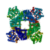 1jpuSC 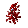 1jq5C C: citing same article ( S: Starting model for refinement |
|---|---|
| Similar structure data |


-Links
 PDBj
PDBj- Assembly
| Deposited unit | 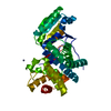
| ||||||||
|---|---|---|---|---|---|---|---|---|---|
| 1 | x 8
| ||||||||
| 2 | 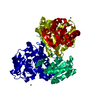
| ||||||||
| Unit cell |
| ||||||||
| Components on special symmetry positions |
| ||||||||
| Details | The biological assembly is an octamer generated from the monomer in the asymmetric unit by the operations: y, -x, z; -x, y, -z; x, -y, -z; y, x, -z; -x, -y, -z; -y, x, z; -y, -x, -z; |
-Components
| #1: Protein | Mass: 39564.996 Da / Num. of mol.: 1 / Mutation: S305C Source method: isolated from a genetically manipulated source Source: (gene. exp.) Geobacillus stearothermophilus (bacteria)Gene: GLDA or GLD / Plasmid: pKK233-2 / Production host: | ||||
|---|---|---|---|---|---|
| #2: Chemical |   Mass: 65.409 Da / Num. of mol.: 2 / Source method: obtained synthetically / Formula: Zn Mass: 65.409 Da / Num. of mol.: 2 / Source method: obtained synthetically / Formula: Zn#3: Chemical | ChemComp-GOL / |   Mass: 92.094 Da / Num. of mol.: 1 / Source method: obtained synthetically / Formula: C3H8O3 Mass: 92.094 Da / Num. of mol.: 1 / Source method: obtained synthetically / Formula: C3H8O3#4: Water | ChemComp-HOH / |  Mass: 18.015 Da / Num. of mol.: 174 / Source method: isolated from a natural source / Formula: H2O Mass: 18.015 Da / Num. of mol.: 174 / Source method: isolated from a natural source / Formula: H2O |
-Experimental details
-Experiment
| Experiment | Method: X-RAY DIFFRACTION / Number of used crystals: 1 |
|---|
- Sample preparation
| Crystal | Density Matthews: 2.65 Å3/Da / Density % sol: 53.56 % |
|---|---|
| Crystal grow | Temperature: 290 K / Method: vapor diffusion, hanging drop Details: Ammonium Sulfate, PEG 400, ZnCl2, VAPOR DIFFUSION, HANGING DROP, temperature 290K |
-Data collection
| Diffraction | Mean temperature: 290 K |
|---|---|
| Diffraction source | Source: SYNCHROTRON / Site: SRS  / Beamline: PX14.2 / Wavelength: 0.97 Å / Beamline: PX14.2 / Wavelength: 0.97 Å |
| Detector | Type: ADSC QUANTUM 4 / Detector: CCD / Date: Nov 29, 1999 |
| Radiation | Protocol: SINGLE WAVELENGTH / Monochromatic (M) / Laue (L): M / Scattering type: x-ray |
| Radiation wavelength | Wavelength: 0.97 Å / Relative weight: 1 |
| Reflection | Resolution: 2.05→10 Å / Num. all: 25033 / Num. obs: 25033 / % possible obs: 93.5 % / Observed criterion σ(F): 0 / Observed criterion σ(I): 0 / Redundancy: 4.2 % / Rmerge(I) obs: 0.081 / Net I/σ(I): 13.2 |
| Reflection shell | Resolution: 2.05→2.1 Å / Rmerge(I) obs: 0.288 / Mean I/σ(I) obs: 2.9 / % possible all: 72 |
- Processing
| Software |
| |||||||||||||||||||||||||||
|---|---|---|---|---|---|---|---|---|---|---|---|---|---|---|---|---|---|---|---|---|---|---|---|---|---|---|---|---|
| Refinement | Method to determine structure: MOLECULAR REPLACEMENT Starting model: PDB ENTRY 1JPU Resolution: 2.05→10 Å / Isotropic thermal model: Isotropic / Cross valid method: THROUGHOUT / σ(F): 1 / Stereochemistry target values: Engh & Huber
| |||||||||||||||||||||||||||
| Displacement parameters | Biso mean: 36.55 Å2 | |||||||||||||||||||||||||||
| Refine analyze |
| |||||||||||||||||||||||||||
| Refinement step | Cycle: LAST / Resolution: 2.05→10 Å
| |||||||||||||||||||||||||||
| Refine LS restraints |
|
