 Movie
Movie Controller

[English] 日本語
 Yorodumi
- PDB-4mca: Crystal Structure of Glycerol Dehydrogenase from Serratia to 1.9A -
+ Open data

- Basic information
| Entry | Database: PDB / ID: 4mca | ||||||
|---|---|---|---|---|---|---|---|
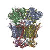
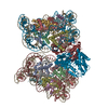
| Title | Crystal Structure of Glycerol Dehydrogenase from Serratia to 1.9A | ||||||
 Components Components | Glycerol dehydrogenase | ||||||
 Keywords Keywords | OXIDOREDUCTASE / Glycerol metabolism | ||||||
| Function / homology | Dehydroquinate synthase-like, alpha domain / Dehydroquinate synthase-like - alpha domain / Rossmann fold - #1970 / Up-down Bundle / Rossmann fold / 3-Layer(aba) Sandwich / Mainly Alpha / Alpha Beta / :  Function and homology information Function and homology information | ||||||
| Biological species |  Serratia plymuthica A30 (bacteria) Serratia plymuthica A30 (bacteria) | ||||||
| Method |  X-RAY DIFFRACTION / SYNCHROTRON / MOLECULAR REPLACEMENT / molecular replacement / Resolution: 1.9 Å X-RAY DIFFRACTION / SYNCHROTRON / MOLECULAR REPLACEMENT / molecular replacement / Resolution: 1.9 Å | ||||||
 Authors Authors | Musille, P.M. / Ortlund, E.A. | ||||||
 Citation Citation | Journal: Acta Crystallogr.,Sect.F / Year: 2014 Title: Structure of glycerol dehydrogenase from Serratia Authors: Musille, P.M. / Ortlund, E.A. | ||||||
| History |
|
- Structure visualization
| Structure viewer | Molecule: MolmilJmol/JSmol |
|---|
- Downloads & links
-Download
| PDBx/mmCIF format | 4mca.cif.gz | 154.9 KB | Display | PDBx/mmCIF format |
|---|---|---|---|---|
| PDB format | pdb4mca.ent.gz | 122.3 KB | Display | PDB format |
| PDBx/mmJSON format | 4mca.json.gz | Tree view | PDBx/mmJSON format | |
| Others |  Other downloads Other downloads |
-Validation report
| Arichive directory | https://data.pdbj.org/pub/pdb/validation_reports/mc/4mcaftp://data.pdbj.org/pub/pdb/validation_reports/mc/4mca | HTTPS FTP |
|---|
-Related structure data
| Similar structure data |
|---|


-Links
 PDBj
PDBj- Assembly
| Deposited unit | 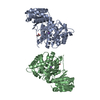
| ||||||||
|---|---|---|---|---|---|---|---|---|---|
| 1 | x 8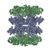
| ||||||||
| 2 | 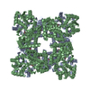
| ||||||||
| 3 | 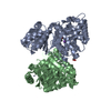
| ||||||||
| Unit cell |
|
-Components
| #1: Protein | Mass: 38802.645 Da / Num. of mol.: 2 / Source method: isolated from a natural source / Source: (natural) Serratia plymuthica A30 (bacteria) / References: UniProt: L0VUH7, glycerol dehydrogenase#2: Chemical | ChemComp-ZN /   Mass: 65.409 Da / Num. of mol.: 4 / Source method: obtained synthetically / Formula: Zn Mass: 65.409 Da / Num. of mol.: 4 / Source method: obtained synthetically / Formula: Zn#3: Chemical | ChemComp-GOL /   Mass: 92.094 Da / Num. of mol.: 4 / Source method: obtained synthetically / Formula: C3H8O3 Mass: 92.094 Da / Num. of mol.: 4 / Source method: obtained synthetically / Formula: C3H8O3#4: Chemical |   Mass: 22.990 Da / Num. of mol.: 2 / Source method: obtained synthetically / Formula: Na Mass: 22.990 Da / Num. of mol.: 2 / Source method: obtained synthetically / Formula: Na#5: Water | ChemComp-HOH / |  Mass: 18.015 Da / Num. of mol.: 365 / Source method: isolated from a natural source / Formula: H2O Mass: 18.015 Da / Num. of mol.: 365 / Source method: isolated from a natural source / Formula: H2O |
|---|
-Experimental details
-Experiment
| Experiment | Method: X-RAY DIFFRACTION / Number of used crystals: 1 |
|---|
- Sample preparation
| Crystal | Density Matthews: 2.89 Å3/Da / Density % sol: 57.44 % |
|---|---|
| Crystal grow | Temperature: 295 K / Method: vapor diffusion, hanging drop / pH: 7.4 Details: 5-10% PEG 3350, 0.2M Calcium Acetate, 4% 2,2,2 trifluoroethanol, 4% glycerol, pH 7.4, VAPOR DIFFUSION, HANGING DROP, temperature 295K |
-Data collection
| Diffraction | Mean temperature: 100 K |
|---|---|
| Diffraction source | Source: SYNCHROTRON / Site: APS  / Beamline: 22-ID / Wavelength: 1 Å / Beamline: 22-ID / Wavelength: 1 Å |
| Detector | Type: MARMOSAIC 300 mm CCD / Detector: CCD / Date: Mar 24, 2013 |
| Radiation | Monochromator: Si 111 CHANNEL / Protocol: SINGLE WAVELENGTH / Monochromatic (M) / Laue (L): M / Scattering type: x-ray |
| Radiation wavelength | Wavelength: 1 Å / Relative weight: 1 |
| Reflection | Resolution: 1.9→50 Å / Num. all: 71425 / Num. obs: 65891 / % possible obs: 91.6 % / Observed criterion σ(F): 0 / Observed criterion σ(I): 0 / Redundancy: 8.7 % / Rmerge(I) obs: 0.099 / Net I/σ(I): 20.2 |
| Reflection shell | Resolution: 1.9→1.97 Å / Redundancy: 4 % / Mean I/σ(I) obs: 2.46 / Num. unique all: 3706 / % possible all: 52.3 |
-Phasing
| Phasing | Method: molecular replacement |
|---|
- Processing
| Software |
| |||||||||||||||||||||||||||||||||||||||||||||||||||||||||||||||||||||||||||
|---|---|---|---|---|---|---|---|---|---|---|---|---|---|---|---|---|---|---|---|---|---|---|---|---|---|---|---|---|---|---|---|---|---|---|---|---|---|---|---|---|---|---|---|---|---|---|---|---|---|---|---|---|---|---|---|---|---|---|---|---|---|---|---|---|---|---|---|---|---|---|---|---|---|---|---|---|
| Refinement | Method to determine structure: MOLECULAR REPLACEMENT / Resolution: 1.9→39.6 Å / Cor.coef. Fo:Fc: 0.96 / Cor.coef. Fo:Fc free: 0.941 / WRfactor Rfree: 0.2009 / WRfactor Rwork: 0.1647 / Occupancy max: 1 / Occupancy min: 0.15 / FOM work R set: 0.8584 / SU B: 3.037 / SU ML: 0.087 / SU R Cruickshank DPI: 0.1353 / SU Rfree: 0.1304 / Cross valid method: THROUGHOUT / σ(F): 0 / ESU R: 0.135 / ESU R Free: 0.13 / Stereochemistry target values: MAXIMUM LIKELIHOOD Details: HYDROGENS HAVE BEEN ADDED IN THE RIDING POSITIONS U VALUES : REFINED INDIVIDUALLY
| |||||||||||||||||||||||||||||||||||||||||||||||||||||||||||||||||||||||||||
| Solvent computation | Ion probe radii: 0.8 Å / Shrinkage radii: 0.8 Å / VDW probe radii: 1.2 Å / Solvent model: MASK | |||||||||||||||||||||||||||||||||||||||||||||||||||||||||||||||||||||||||||
| Displacement parameters | Biso max: 101.78 Å2 / Biso mean: 30.0239 Å2 / Biso min: 11.41 Å2
| |||||||||||||||||||||||||||||||||||||||||||||||||||||||||||||||||||||||||||
| Refinement step | Cycle: LAST / Resolution: 1.9→39.6 Å
| |||||||||||||||||||||||||||||||||||||||||||||||||||||||||||||||||||||||||||
| Refine LS restraints |
| |||||||||||||||||||||||||||||||||||||||||||||||||||||||||||||||||||||||||||
| LS refinement shell | Resolution: 1.898→1.948 Å / Total num. of bins used: 20
|
