 Movie
Movie Controller


+ Open data

- Basic information
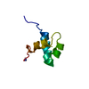
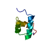
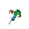
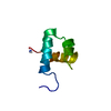
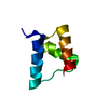
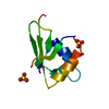
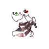
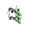
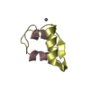
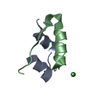
| Entry | Database: PDB / ID: 1gv5 | ||||||
|---|---|---|---|---|---|---|---|
| Title | CRYSTAL STRUCTURE OF C-MYB R2 | ||||||
 Components Components | MYB PROTO-ONCOGENE PROTEIN | ||||||
 Keywords Keywords | TRANSCRIPTION / TRANSCRIPTION REGULATION / MYB / C-MYB / DNA BINDING / ION BINDI PROTO-ONCOGENE / NUCLEAR PROTEIN | ||||||
| Function / homology |  Function and homology information Function and homology informationpositive regulation of testosterone secretion / myeloid cell development / positive regulation of hepatic stellate cell proliferation / skeletal muscle cell proliferation / negative regulation of hematopoietic progenitor cell differentiation / positive regulation of hepatic stellate cell activation / positive regulation of transforming growth factor beta production / embryonic digestive tract development / myeloid cell differentiation / cellular response to interleukin-6 ...positive regulation of testosterone secretion / myeloid cell development / positive regulation of hepatic stellate cell proliferation / skeletal muscle cell proliferation / negative regulation of hematopoietic progenitor cell differentiation / positive regulation of hepatic stellate cell activation / positive regulation of transforming growth factor beta production / embryonic digestive tract development / myeloid cell differentiation / cellular response to interleukin-6 / T-helper 2 cell differentiation / stem cell division / WD40-repeat domain binding / positive regulation of collagen biosynthetic process / homeostasis of number of cells / positive regulation of glial cell proliferation / spleen development / negative regulation of megakaryocyte differentiation / cellular response to retinoic acid / positive regulation of smooth muscle cell proliferation / B cell differentiation / thymus development / response to ischemia / cellular response to leukemia inhibitory factor / erythrocyte differentiation / G1/S transition of mitotic cell cycle / positive regulation of miRNA transcription / RNA polymerase II transcription regulator complex / cellular response to hydrogen peroxide / calcium ion transport / positive regulation of neuron apoptotic process / mitotic cell cycle / regulation of gene expression / DNA-binding transcription activator activity, RNA polymerase II-specific / in utero embryonic development / DNA-binding transcription factor activity, RNA polymerase II-specific / response to hypoxia / RNA polymerase II cis-regulatory region sequence-specific DNA binding / DNA-binding transcription factor activity / negative regulation of DNA-templated transcription / regulation of DNA-templated transcription / positive regulation of DNA-templated transcription / negative regulation of transcription by RNA polymerase II / positive regulation of transcription by RNA polymerase II / DNA binding / nucleoplasm / nucleus / cytosol Similarity search - Function | ||||||
| Biological species |  | ||||||
| Method |  X-RAY DIFFRACTION / MOLECULAR REPLACEMENT / Resolution: 1.58 Å X-RAY DIFFRACTION / MOLECULAR REPLACEMENT / Resolution: 1.58 Å | ||||||
 Authors Authors | Tahirov, T.H. / Ogata, K. | ||||||
 Citation Citation | Journal: To be Published Title: Crystal Structure of C-Myb DNA-Binding Domain: Specific Na+ Binding and Correlation with NMR Structure Authors: Tahirov, T.H. / Morii, H. / Uedaira, H. / Sasaki, M. / Sarai, A. / Adachi, S. / Park, S.Y. / Kamiya, N. / Ogata, K. #1: Journal: Cell (Cambridge,Mass.) / Year: 2002Title: Mechanism of C-Myb-C/Ebpbeta Cooperation from Separated Sites on a Promoter Authors: Tahirov, T.H. / Sato, K. / Ichikawa-Iwata, E. / Sasaki, M. / Inoue-Bungo, T. / Shiina, M. / Kimura, K. / Takata, S. / Fujikawa, A. / Morii, H. / Kumasaka, T. / Yamamoto, M. / Ishii, S. / Ogata, K. #2: Journal: Acta Crystallogr.,Sect.D / Year: 1999 Title: Crystallization and Preliminary X-Ray Analysis of Wild Type and V103L Mutant Myb R2 DNA-Binding Domain Authors: Tahirov, T.H. / Morii, H. / Uedaira, H. / Sarai, A. / Ogata, K. | ||||||
| History |
|
- Structure visualization
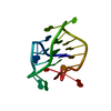
| Structure viewer | Molecule: MolmilJmol/JSmol |
|---|
- Downloads & links
-Download
| PDBx/mmCIF format | 1gv5.cif.gz | 25.5 KB | Display | PDBx/mmCIF format |
|---|---|---|---|---|
| PDB format | pdb1gv5.ent.gz | 15.7 KB | Display | PDB format |
| PDBx/mmJSON format | 1gv5.json.gz | Tree view | PDBx/mmJSON format | |
| Others |  Other downloads Other downloads |
-Validation report
| Arichive directory | https://data.pdbj.org/pub/pdb/validation_reports/gv/1gv5ftp://data.pdbj.org/pub/pdb/validation_reports/gv/1gv5 | HTTPS FTP |
|---|
-Related structure data
| Related structure data |  1guuC 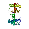 1gv2C 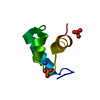 1gvdC 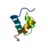 1mbgS C: citing same article ( S: Starting model for refinement |
|---|---|
| Similar structure data |


-Links
 PDBj
PDBj
- Assembly
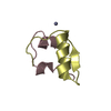
| Deposited unit | 
| ||||||||
|---|---|---|---|---|---|---|---|---|---|
| 1 |
| ||||||||
| Unit cell |
|
-Components
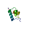
| #1: Protein | Mass: 6302.409 Da / Num. of mol.: 1 / Fragment: R2, RESIDUES 90-141 / Source method: obtained synthetically / Source: (synth.) |
|---|---|
| #2: Chemical | ChemComp-NA /   Mass: 22.990 Da / Num. of mol.: 1 / Source method: obtained synthetically / Formula: Na Mass: 22.990 Da / Num. of mol.: 1 / Source method: obtained synthetically / Formula: Na |
| #3: Water | ChemComp-HOH /  Mass: 18.015 Da / Num. of mol.: 50 / Source method: isolated from a natural source / Formula: H2O Mass: 18.015 Da / Num. of mol.: 50 / Source method: isolated from a natural source / Formula: H2O |
-Experimental details
-Experiment
| Experiment | Method: X-RAY DIFFRACTION / Number of used crystals: 1 |
|---|
- Sample preparation
| Crystal | Density Matthews: 2.26 Å3/Da / Density % sol: 45.9 % |
|---|---|
| Crystal grow | Temperature: 297 K / pH: 6.8 Details: 1.65 M SODIUM CITRATE PH 6.8, PROTEIN CONCENTRATION 10 MG/ML PLUS 10 MM DTT, TEMPERATURE 297 K |
-Data collection
| Diffraction | Mean temperature: 293 K |
|---|---|
| Diffraction source | Source: ROTATING ANODE / Type: MAC SCIENCE M06XHF22 / Wavelength: 1.5418 |
| Detector | Type: MAC Science DIP-2030 / Detector: IMAGE PLATE / Date: Oct 29, 1997 / Details: MAC SCIENCE DOUBLE MIRROR |
| Radiation | Monochromator: 0.15 MM NICKEL FILTER / Protocol: SINGLE WAVELENGTH / Monochromatic (M) / Laue (L): M / Scattering type: x-ray |
| Radiation wavelength | Wavelength: 1.5418 Å / Relative weight: 1 |
| Reflection | Resolution: 1.58→20 Å / Num. obs: 8207 / % possible obs: 99.7 % / Observed criterion σ(I): 0 / Redundancy: 11.16 % / Biso Wilson estimate: 17.1 Å2 / Rmerge(I) obs: 0.059 / Net I/σ(I): 42.736 |
| Reflection shell | Resolution: 1.58→1.64 Å / Redundancy: 6.17 % / Rmerge(I) obs: 0.329 / Mean I/σ(I) obs: 5.285 / % possible all: 98.9 |
- Processing
| Software |
| ||||||||||||||||||||||||||||||||||||||||||||||||||||||||||||||||||||||||||||||||
|---|---|---|---|---|---|---|---|---|---|---|---|---|---|---|---|---|---|---|---|---|---|---|---|---|---|---|---|---|---|---|---|---|---|---|---|---|---|---|---|---|---|---|---|---|---|---|---|---|---|---|---|---|---|---|---|---|---|---|---|---|---|---|---|---|---|---|---|---|---|---|---|---|---|---|---|---|---|---|---|---|---|
| Refinement | Method to determine structure: MOLECULAR REPLACEMENT Starting model: PDB ENTRY 1MBG Resolution: 1.58→14.26 Å / Rfactor Rfree error: 0.009 / Data cutoff high absF: 408120.47 / Isotropic thermal model: RESTRAINED / Cross valid method: THROUGHOUT / σ(F): 0
| ||||||||||||||||||||||||||||||||||||||||||||||||||||||||||||||||||||||||||||||||
| Solvent computation | Solvent model: FLAT MODEL / Bsol: 90.0897 Å2 / ksol: 0.574584 e/Å3 | ||||||||||||||||||||||||||||||||||||||||||||||||||||||||||||||||||||||||||||||||
| Displacement parameters | Biso mean: 19.1 Å2
| ||||||||||||||||||||||||||||||||||||||||||||||||||||||||||||||||||||||||||||||||
| Refine analyze |
| ||||||||||||||||||||||||||||||||||||||||||||||||||||||||||||||||||||||||||||||||
| Refinement step | Cycle: LAST / Resolution: 1.58→14.26 Å
| ||||||||||||||||||||||||||||||||||||||||||||||||||||||||||||||||||||||||||||||||
| Refine LS restraints |
| ||||||||||||||||||||||||||||||||||||||||||||||||||||||||||||||||||||||||||||||||
| LS refinement shell | Resolution: 1.58→1.68 Å / Rfactor Rfree error: 0.031 / Total num. of bins used: 6
| ||||||||||||||||||||||||||||||||||||||||||||||||||||||||||||||||||||||||||||||||
| Xplor file |
|
