 Movie
Movie Controller

[English] 日本語
 Yorodumi
- PDB-1mse: SOLUTION STRUCTURE OF A SPECIFIC DNA COMPLEX OF THE MYB DNA-BINDI... -
+ Open data

- Basic information
| Entry | Database: PDB / ID: 1mse | ||||||
|---|---|---|---|---|---|---|---|
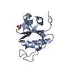
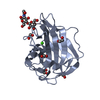
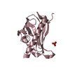
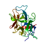
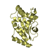
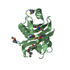
| Title | SOLUTION STRUCTURE OF A SPECIFIC DNA COMPLEX OF THE MYB DNA-BINDING DOMAIN WITH COOPERATIVE RECOGNITION HELICES | ||||||
 Components Components |
| ||||||
 Keywords Keywords | DNA BINDING PROTEIN/DNA / DNA / DOUBLE HELIX / C-MYB DNA-BINDING DOMAIN / PROTOONCOGENE PRODUCT / DNA BINDING PROTEIN-DNA COMPLEX | ||||||
| Function / homology |  Function and homology information Function and homology informationpositive regulation of testosterone secretion / myeloid cell development / positive regulation of hepatic stellate cell proliferation / skeletal muscle cell proliferation / negative regulation of hematopoietic progenitor cell differentiation / positive regulation of hepatic stellate cell activation / positive regulation of transforming growth factor beta production / embryonic digestive tract development / myeloid cell differentiation / cellular response to interleukin-6 ...positive regulation of testosterone secretion / myeloid cell development / positive regulation of hepatic stellate cell proliferation / skeletal muscle cell proliferation / negative regulation of hematopoietic progenitor cell differentiation / positive regulation of hepatic stellate cell activation / positive regulation of transforming growth factor beta production / embryonic digestive tract development / myeloid cell differentiation / cellular response to interleukin-6 / T-helper 2 cell differentiation / stem cell division / WD40-repeat domain binding / positive regulation of collagen biosynthetic process / homeostasis of number of cells / positive regulation of glial cell proliferation / spleen development / negative regulation of megakaryocyte differentiation / cellular response to retinoic acid / positive regulation of smooth muscle cell proliferation / B cell differentiation / thymus development / response to ischemia / cellular response to leukemia inhibitory factor / erythrocyte differentiation / G1/S transition of mitotic cell cycle / positive regulation of miRNA transcription / RNA polymerase II transcription regulator complex / cellular response to hydrogen peroxide / calcium ion transport / positive regulation of neuron apoptotic process / mitotic cell cycle / regulation of gene expression / DNA-binding transcription activator activity, RNA polymerase II-specific / in utero embryonic development / DNA-binding transcription factor activity, RNA polymerase II-specific / response to hypoxia / RNA polymerase II cis-regulatory region sequence-specific DNA binding / DNA-binding transcription factor activity / negative regulation of DNA-templated transcription / regulation of DNA-templated transcription / positive regulation of DNA-templated transcription / negative regulation of transcription by RNA polymerase II / positive regulation of transcription by RNA polymerase II / DNA binding / nucleoplasm / nucleus / cytosol Similarity search - Function | ||||||
| Biological species |  | ||||||
| Method | SOLUTION NMR / ENERGY MINIMIZATION | ||||||
 Authors Authors | Ogata, K. / Morikawa, S. / Nakamura, H. / Sekikawa, A. / Inoue, T. / Kanai, H. / Sarai, A. / Ishii, S. / Nishimura, Y. | ||||||
 Citation Citation | Journal: Cell(Cambridge,Mass.) / Year: 1994 Title: Solution structure of a specific DNA complex of the Myb DNA-binding domain with cooperative recognition helices. Authors: Ogata, K. / Morikawa, S. / Nakamura, H. / Sekikawa, A. / Inoue, T. / Kanai, H. / Sarai, A. / Ishii, S. / Nishimura, Y. #1: Journal: Proc.Natl.Acad.Sci.USA / Year: 1992Title: Solution Structure of a DNA-Binding Unit of Myb: A Helix-Turn-Helix-Related Motif with Conserved Tryptophans Forming a Hydrophobic Core Authors: Ogata, K. / Hojo, H. / Aimoto, S. / Nakai, T. / Nakamura, H. / Sarai, A. / Ishii, S. / Nishimura, Y. | ||||||
| History |
|
- Structure visualization
| Structure viewer | Molecule: MolmilJmol/JSmol |
|---|
- Downloads & links
-Download
| PDBx/mmCIF format | 1mse.cif.gz | 65.9 KB | Display | PDBx/mmCIF format |
|---|---|---|---|---|
| PDB format | pdb1mse.ent.gz | 46 KB | Display | PDB format |
| PDBx/mmJSON format | 1mse.json.gz | Tree view | PDBx/mmJSON format | |
| Others |  Other downloads Other downloads |
-Validation report
| Arichive directory | https://data.pdbj.org/pub/pdb/validation_reports/ms/1mseftp://data.pdbj.org/pub/pdb/validation_reports/ms/1mse | HTTPS FTP |
|---|
-Related structure data


-Links
 PDBj
PDBj


- Assembly
| Deposited unit | 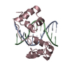
| |||||||||
|---|---|---|---|---|---|---|---|---|---|---|
| 1 |
| |||||||||
| NMR ensembles |
|
-Components
| #1: DNA chain | Mass: 4811.158 Da / Num. of mol.: 1 / Source method: obtained synthetically / Details: CHEMICALLY SYNTHESIZED / Keywords: DEOXYRIBONUCLEIC ACID |
|---|---|
| #2: DNA chain | Mass: 4984.237 Da / Num. of mol.: 1 / Source method: obtained synthetically / Details: CHEMICALLY SYNTHESIZED / Keywords: DEOXYRIBONUCLEIC ACID |
| #3: Protein | Mass: 12676.775 Da / Num. of mol.: 1 Source method: isolated from a genetically manipulated source Source: (gene. exp.)  Keywords: POLYPEPTIDE / References: UniProt: P06876 Keywords: POLYPEPTIDE / References: UniProt: P06876 |
-Experimental details
-Experiment
| Experiment | Method: SOLUTION NMR |
|---|---|
| NMR details | Text: THE STRUCTURES OF N- AND C-TERMINI OF THE PEPTIDE CHAIN (MET 89 - PRO 94 AND ASN 186 - VAL 193) WERE DISORDERED AMONG THE 25 STRUCTURES. THE ORIENTATION OF THE LAST 5 BASE PAIRS OF THE DNA IS ...Text: THE STRUCTURES OF N- AND C-TERMINI OF THE PEPTIDE CHAIN (MET 89 - PRO 94 AND ASN 186 - VAL 193) WERE DISORDERED AMONG THE 25 STRUCTURES. THE ORIENTATION OF THE LAST 5 BASE PAIRS OF THE DNA IS POORLY DEFINED WITH RESPECT TO THE CORE OF THE COMPLEX. CONSEQUENTLY, ONLY THE COORDINATES OF BASE PAIRS 1 - 11 OF DNA HAVE BEEN DEPOSITED TOGETHER WITH THOSE OF THE PROTEIN IN THE COMPLEX. |
- Sample preparation
| Sample conditions | pH: 6.8 / Temperature: 310 K |
|---|---|
| Crystal grow | *PLUS Method: other / Details: NMR |
- Processing
| Software | Name:  AMBER / Classification: refinement AMBER / Classification: refinement | |||||||||
|---|---|---|---|---|---|---|---|---|---|---|
| NMR software |
| |||||||||
| Refinement | Method: ENERGY MINIMIZATION / Software ordinal: 1 Details: RMSD BOND DISTANCES 0.006 ANGSTROMS RMSD BOND ANGLES 0.857 DEGREES RMSD CHIRAL CENTERS 0.0664 ANGSTROMS NUMBER OF ATOMS USED IN REFINEMENT. NUMBER OF PROTEIN ATOMS 1815 NUMBER OF NUCLEIC ...Details: RMSD BOND DISTANCES 0.006 ANGSTROMS RMSD BOND ANGLES 0.857 DEGREES RMSD CHIRAL CENTERS 0.0664 ANGSTROMS NUMBER OF ATOMS USED IN REFINEMENT. NUMBER OF PROTEIN ATOMS 1815 NUMBER OF NUCLEIC ACID ATOMS 698 AMBER FORCE FIELD | |||||||||
| NMR constraints | NOE constraints total: 1732 / Hydrogen bond constraints total count: 350 | |||||||||
| NMR ensemble | Conformer selection criteria: structures with the least restraint violations,structures with the lowest energy Conformers calculated total number: 25 / Conformers submitted total number: 1 |
