[English] 日本語
Yorodumi
- PDB-1gvd: CRYSTAL STRUCTURE OF C-MYB R2 V103L MUTANT -

+
Open data


ID or keywords:

Loading...

-
Basic information

Entry
Database: PDB / ID: 1gvd
TitleCRYSTAL STRUCTURE OF C-MYB R2 V103L MUTANT
ComponentsMYB PROTO-ONCOGENE PROTEIN
KeywordsTRANSCRIPTION / TRANSCRIPTION REGULATION / MYB / C-MYB / DNA BINDING / ION BINDING / PROTO-ONCOGENE / NUCLEAR PROTEIN
Function / homology
Function and homology information


positive regulation of testosterone secretion / myeloid cell development / positive regulation of hepatic stellate cell proliferation / positive regulation of hepatic stellate cell activation / negative regulation of hematopoietic progenitor cell differentiation / positive regulation of transforming growth factor beta production / embryonic digestive tract development / stem cell division / myeloid cell differentiation / cellular response to interleukin-6 ...positive regulation of testosterone secretion / myeloid cell development / positive regulation of hepatic stellate cell proliferation / positive regulation of hepatic stellate cell activation / negative regulation of hematopoietic progenitor cell differentiation / positive regulation of transforming growth factor beta production / embryonic digestive tract development / stem cell division / myeloid cell differentiation / cellular response to interleukin-6 / T-helper 2 cell differentiation / WD40-repeat domain binding / homeostasis of number of cells / positive regulation of collagen biosynthetic process / negative regulation of megakaryocyte differentiation / positive regulation of glial cell proliferation / spleen development / cellular response to leukemia inhibitory factor / erythrocyte differentiation / B cell differentiation / thymus development / positive regulation of smooth muscle cell proliferation / G1/S transition of mitotic cell cycle / positive regulation of miRNA transcription / RNA polymerase II transcription regulator complex / positive regulation of neuron apoptotic process / calcium ion transport / mitotic cell cycle / regulation of gene expression / DNA-binding transcription activator activity, RNA polymerase II-specific / in utero embryonic development / DNA-binding transcription factor activity, RNA polymerase II-specific / RNA polymerase II cis-regulatory region sequence-specific DNA binding / DNA-binding transcription factor activity / negative regulation of DNA-templated transcription / regulation of DNA-templated transcription / positive regulation of DNA-templated transcription / negative regulation of transcription by RNA polymerase II / positive regulation of transcription by RNA polymerase II / DNA binding / nucleoplasm / nucleus / cytosol
Similarity search - Function
Transcription regulator Wos2-domain / LMSTEN motif / C-myb, C-terminal / C-myb, C-terminal / Myb-type HTH DNA-binding domain profile. / Myb domain / Myb-like DNA-binding domain / SANT SWI3, ADA2, N-CoR and TFIIIB'' DNA-binding domains / SANT/Myb domain / Homeodomain-like ...Transcription regulator Wos2-domain / LMSTEN motif / C-myb, C-terminal / C-myb, C-terminal / Myb-type HTH DNA-binding domain profile. / Myb domain / Myb-like DNA-binding domain / SANT SWI3, ADA2, N-CoR and TFIIIB'' DNA-binding domains / SANT/Myb domain / Homeodomain-like / Homeobox-like domain superfamily / Arc Repressor Mutant, subunit A / Orthogonal Bundle / Mainly Alpha
Similarity search - Domain/homology
AMMONIUM ION / Transcriptional activator Myb
Similarity search - Component
Biological speciesMUS MUSCULUS (house mouse)
MethodX-RAY DIFFRACTION / MOLECULAR REPLACEMENT / Resolution: 1.45 Å
AuthorsTahirov, T.H. / Ogata, K.
Citation
Journal: To be Published
Title: Crystal Structure of C-Myb DNA-Binding Domain: Specific Na+ Binding and Correlation with NMR Structure
Authors: Tahirov, T.H. / Morii, H. / Uedaira, H. / Sasaki, M. / Sarai, A. / Adachi, S. / Park, S.Y. / Kamiya, N. / Ogata, K.
#1: Journal: Cell (Cambridge,Mass.) / Year: 2002
Title: Mechanism of C-Myb-C/Ebpbeta Cooperation from Separated Sites on a Promoter
Authors: Tahirov, T.H. / Sato, K. / Ichikawa-Iwata, E. / Sasaki, M. / Inoue-Bungo, T. / Shiina, M. / Kimura, K. / Takata, S. / Fujikawa, A. / Morii, H. / Kumasaka, T. / Yamamoto, M. / Ishii, S. / Ogata, K.
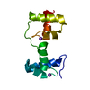
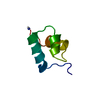
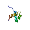
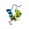
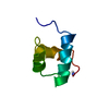
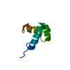
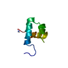
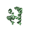
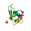
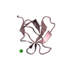
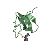
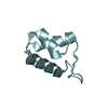
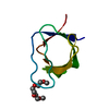
#2: Journal: Acta Crystallogr.,Sect.D / Year: 1999
Title: Crystallization and Preliminary X-Ray Analysis of Wild Type and V103L Mutant Myb R2 DNA-Binding Domain
Authors: Tahirov, T.H. / Morii, H. / Uedaira, H. / Sarai, A. / Ogata, K.
History
DepositionFeb 8, 2002Deposition site: PDBE / Processing site: PDBE
Revision 1.0Jul 3, 2003Provider: repository / Type: Initial release
Revision 1.1May 8, 2011Group: Version format compliance
Revision 1.2Jul 13, 2011Group: Version format compliance
Revision 1.3May 8, 2019Group: Data collection / Experimental preparation / Other
Category: exptl_crystal_grow / pdbx_database_proc / pdbx_database_status
Item: _exptl_crystal_grow.temp / _pdbx_database_status.recvd_author_approval
Revision 1.4Dec 13, 2023Group: Data collection / Database references ...Data collection / Database references / Other / Refinement description
Category: chem_comp_atom / chem_comp_bond ...chem_comp_atom / chem_comp_bond / database_2 / pdbx_database_status / pdbx_initial_refinement_model
Item: _database_2.pdbx_DOI / _database_2.pdbx_database_accession / _pdbx_database_status.status_code_sf

-
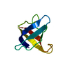
Structure visualization

Structure viewerMolecule:
MolmilJmol/JSmol

Downloads & links

-
Assembly

Deposited unit
A: MYB PROTO-ONCOGENE PROTEIN
hetero molecules


Theoretical massNumber of molelcules
Total (without water)6,5274
Polymers6,3161
Non-polymers2103
Water91951
1


  • Idetical with deposited unit
  • defined by author&software
TypeNameSymmetry operationNumber
identity operation1_555x,y,z1
MethodPQS
Unit cell
Length a, b, c (Å)28.951, 40.114, 48.807
Angle α, β, γ (deg.)90.00, 90.00, 90.00
Int Tables number19
Space group name H-MP212121

-
Components

#1: Protein MYB PROTO-ONCOGENE PROTEIN


Mass: 6316.437 Da / Num. of mol.: 1 / Fragment: R2, RESIDUES 90-141 / Mutation: YES / Source method: obtained synthetically / Source: (synth.) MUS MUSCULUS (house mouse) / References: UniProt: P06876
#2: Chemical ChemComp-NH4 / AMMONIUM ION / Ammonium


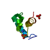
Mass: 18.038 Da / Num. of mol.: 1 / Source method: obtained synthetically / Formula: H4N
#3: Chemical ChemComp-SO4 / SULFATE ION / Sulfate


Mass: 96.063 Da / Num. of mol.: 2 / Source method: obtained synthetically / Formula: SO4
#4: Water ChemComp-HOH / water / Water


Mass: 18.015 Da / Num. of mol.: 51 / Source method: isolated from a natural source / Formula: H2O
Compound detailsENGINEERED MUTATION VAL 103 LEU

-
Experimental details

-
Experiment

ExperimentMethod: X-RAY DIFFRACTION / Number of used crystals: 1

-
Sample preparation

CrystalDensity Matthews: 2.24 Å3/Da / Density % sol: 45.9 %
Crystal growTemperature: 297 K / pH: 6.8
Details: 3.15 M AMMONIUM SULFATE IN 0.05 M MES BUFFER AT PH 6.8, PROTEIN CONCENTRATION 10 MG/ML PLUS 10 MM DTT, TEMPERATURE 297 K

-
Data collection

DiffractionMean temperature: 293 K
Diffraction sourceSource: ROTATING ANODE / Type: MAC SCIENCE M06XHF22 / Wavelength: 1.5418
DetectorType: MAC Science DIP-2030 / Detector: IMAGE PLATE / Date: Jul 16, 1997 / Details: MAC SCIENCE DOUBLE MIRROR
RadiationMonochromator: 0.15 MM NICKEL FILTER / Protocol: SINGLE WAVELENGTH / Monochromatic (M) / Laue (L): M / Scattering type: x-ray
Radiation wavelengthWavelength: 1.5418 Å / Relative weight: 1
ReflectionResolution: 1.45→20 Å / Num. obs: 10332 / % possible obs: 98 % / Observed criterion σ(I): 0 / Redundancy: 8.657 % / Biso Wilson estimate: 14.4 Å2 / Rmerge(I) obs: 0.063 / Net I/σ(I): 38.204
Reflection shellResolution: 1.45→1.5 Å / Redundancy: 6.07 % / Rmerge(I) obs: 0.2 / Mean I/σ(I) obs: 8.351 / % possible all: 95.2

-
Processing

Software
NameVersionClassification
CNS1refinement
DENZOdata reduction
SCALEPACKdata scaling
X-PLORphasing
RefinementMethod to determine structure: MOLECULAR REPLACEMENT
Starting model: PDB ENTRY 1GV5
Resolution: 1.45→12.45 Å / Rfactor Rfree error: 0.008 / Data cutoff high absF: 731165.04 / Isotropic thermal model: RESTRAINED / Cross valid method: THROUGHOUT / σ(F): 0
RfactorNum. reflection% reflectionSelection details
Rfree0.195 543 5.3 %RANDOM
Rwork0.18 ---
obs0.18 10307 97.9 %-
Solvent computationSolvent model: FLAT MODEL / Bsol: 80.5097 Å2 / ksol: 0.550446 e/Å3
Displacement parametersBiso mean: 16.5 Å2
Baniso -1Baniso -2Baniso -3
1-0.87 Å20 Å20 Å2
2---1.24 Å20 Å2
3---0.37 Å2
Refine analyze
FreeObs
Luzzati coordinate error0.16 Å0.15 Å
Luzzati d res low-20 Å
Luzzati sigma a0.07 Å0.06 Å
Refinement stepCycle: LAST / Resolution: 1.45→12.45 Å
ProteinNucleic acidLigandSolventTotal
Num. atoms446 0 11 51 508
Refine LS restraints
Refine-IDTypeDev idealDev ideal target
X-RAY DIFFRACTIONc_bond_d0.003
X-RAY DIFFRACTIONc_bond_d_na
X-RAY DIFFRACTIONc_bond_d_prot
X-RAY DIFFRACTIONc_angle_d
X-RAY DIFFRACTIONc_angle_d_na
X-RAY DIFFRACTIONc_angle_d_prot
X-RAY DIFFRACTIONc_angle_deg1.1
X-RAY DIFFRACTIONc_angle_deg_na
X-RAY DIFFRACTIONc_angle_deg_prot
X-RAY DIFFRACTIONc_dihedral_angle_d18.3
X-RAY DIFFRACTIONc_dihedral_angle_d_na
X-RAY DIFFRACTIONc_dihedral_angle_d_prot
X-RAY DIFFRACTIONc_improper_angle_d0.72
X-RAY DIFFRACTIONc_improper_angle_d_na
X-RAY DIFFRACTIONc_improper_angle_d_prot
X-RAY DIFFRACTIONc_mcbond_it1.941.5
X-RAY DIFFRACTIONc_mcangle_it2.82
X-RAY DIFFRACTIONc_scbond_it3.212
X-RAY DIFFRACTIONc_scangle_it4.742.5
LS refinement shellResolution: 1.45→1.54 Å / Rfactor Rfree error: 0.032 / Total num. of bins used: 6
RfactorNum. reflection% reflection
Rfree0.253 61 3.8 %
Rwork0.236 1543 -
obs--94 %
Xplor file
Refine-IDSerial noParam fileTopol file
X-RAY DIFFRACTION1PROTEIN_REP.PARAMPROTEIN.TOP
X-RAY DIFFRACTION2ION.PARAMION.TOP
X-RAY DIFFRACTION3WATER_REP.PARAMWATER.TOP

+
About Yorodumi

-
News

-
Feb 9, 2022. New format data for meta-information of EMDB entries

New format data for meta-information of EMDB entries

  • Version 3 of the EMDB header file is now the official format.
  • The previous official version 1.9 will be removed from the archive.

Related info.:EMDB header

External links:wwPDB to switch to version 3 of the EMDB data model

-
Aug 12, 2020. Covid-19 info

Covid-19 info

URL: https://pdbj.org/emnavi/covid19.php

New page: Covid-19 featured information page in EM Navigator.

Related info.:Covid-19 info / Mar 5, 2020. Novel coronavirus structure data

+
Mar 5, 2020. Novel coronavirus structure data

Novel coronavirus structure data

Related info.:Yorodumi Speices / Aug 12, 2020. Covid-19 info

External links:COVID-19 featured content - PDBj / Molecule of the Month (242):Coronavirus Proteases

+
Jan 31, 2019. EMDB accession codes are about to change! (news from PDBe EMDB page)

EMDB accession codes are about to change! (news from PDBe EMDB page)

  • The allocation of 4 digits for EMDB accession codes will soon come to an end. Whilst these codes will remain in use, new EMDB accession codes will include an additional digit and will expand incrementally as the available range of codes is exhausted. The current 4-digit format prefixed with “EMD-” (i.e. EMD-XXXX) will advance to a 5-digit format (i.e. EMD-XXXXX), and so on. It is currently estimated that the 4-digit codes will be depleted around Spring 2019, at which point the 5-digit format will come into force.
  • The EM Navigator/Yorodumi systems omit the EMD- prefix.

Related info.:Q: What is EMD? / ID/Accession-code notation in Yorodumi/EM Navigator

External links:EMDB Accession Codes are Changing Soon! / Contact to PDBj

+
Jul 12, 2017. Major update of PDB

Major update of PDB

  • wwPDB released updated PDB data conforming to the new PDBx/mmCIF dictionary.
  • This is a major update changing the version number from 4 to 5, and with Remediation, in which all the entries are updated.
  • In this update, many items about electron microscopy experimental information are reorganized (e.g. em_software).
  • Now, EM Navigator and Yorodumi are based on the updated data.

External links:wwPDB Remediation / Enriched Model Files Conforming to OneDep Data Standards Now Available in the PDB FTP Archive

-
Yorodumi

Thousand views of thousand structures

  • Yorodumi is a browser for structure data from EMDB, PDB, SASBDB, etc.
  • This page is also the successor to EM Navigator detail page, and also detail information page/front-end page for Omokage search.
  • The word "yorodu" (or yorozu) is an old Japanese word meaning "ten thousand". "mi" (miru) is to see.

Related info.:EMDB / PDB / SASBDB / Comparison of 3 databanks / Yorodumi Search / Aug 31, 2016. New EM Navigator & Yorodumi / Yorodumi Papers / Jmol/JSmol / Function and homology information / Changes in new EM Navigator and Yorodumi

Read more