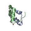
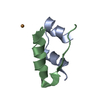
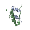
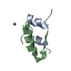
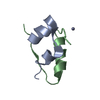
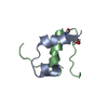
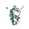
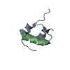
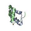
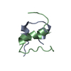
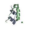
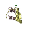
- PDB-4m4m: The structure of Ni T6 bovine insulin -
+
Open data
ID or keywords:
Loading...
-
Basic information
Entry
Database: PDB / ID: 4m4m
Title
The structure of Ni T6 bovine insulin
Components
(Insulin) x 2
Keywords
HORMONE / Nickel binding
Function / homology
Function and homology information
estradiol secretion / positive regulation of blood circulation / negative regulation of lactation / glucose import in response to insulin stimulus / positive regulation of cell maturation / response to L-arginine / positive regulation of mammary gland epithelial cell proliferation / positive regulation of lactation / response to butyrate / negative regulation of appetite ...estradiol secretion / positive regulation of blood circulation / negative regulation of lactation / glucose import in response to insulin stimulus / positive regulation of cell maturation / response to L-arginine / positive regulation of mammary gland epithelial cell proliferation / positive regulation of lactation / response to butyrate / negative regulation of appetite / feeding behavior / response to growth hormone / positive regulation of peptide hormone secretion / response to food / positive regulation of Rho protein signal transduction / protein secretion / negative regulation of lipid catabolic process / response to glucose / insulin receptor binding / positive regulation of protein secretion / hormone activity / response to nutrient levels / glucose metabolic process / positive regulation of insulin secretion / glucose homeostasis / response to heat / positive regulation of phosphatidylinositol 3-kinase/protein kinase B signal transduction / positive regulation of gene expression / negative regulation of apoptotic process / : / identical protein binding Similarity search - Function
Insulin / Insulin family / Insulin-like / Insulin/IGF/Relaxin family / Insulin / insulin-like growth factor / relaxin family. / Insulin, conserved site / Insulin family signature. / Insulin-like superfamily Similarity search - Domain/homology
In the structure databanks used in Yorodumi, some data are registered as the other names, "COVID-19 virus" and "2019-nCoV". Here are the details of the virus and the list of structure data.
Jan 31, 2019. EMDB accession codes are about to change! (news from PDBe EMDB page)
EMDB accession codes are about to change! (news from PDBe EMDB page)
The allocation of 4 digits for EMDB accession codes will soon come to an end. Whilst these codes will remain in use, new EMDB accession codes will include an additional digit and will expand incrementally as the available range of codes is exhausted. The current 4-digit format prefixed with “EMD-” (i.e. EMD-XXXX) will advance to a 5-digit format (i.e. EMD-XXXXX), and so on. It is currently estimated that the 4-digit codes will be depleted around Spring 2019, at which point the 5-digit format will come into force.
The EM Navigator/Yorodumi systems omit the EMD- prefix.
Related info.:Q: What is EMD? / ID/Accession-code notation in Yorodumi/EM Navigator
Yorodumi is a browser for structure data from EMDB, PDB, SASBDB, etc.
This page is also the successor to EM Navigator detail page, and also detail information page/front-end page for Omokage search.
The word "yorodu" (or yorozu) is an old Japanese word meaning "ten thousand". "mi" (miru) is to see.
Related info.:EMDB / PDB / SASBDB / Comparison of 3 databanks / Yorodumi Search / Aug 31, 2016. New EM Navigator & Yorodumi / Yorodumi Papers / Jmol/JSmol / Function and homology information / Changes in new EM Navigator and Yorodumi
 Movie
Movie Controller


 Open data
 Basic information
 Components
 Keywords
 Function and homology information
 X-RAY DIFFRACTION /
  Authors
 Citation
 Structure visualization
 Downloads & links
 Other downloads
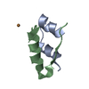


 PDBj
PDBj


 Assembly
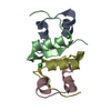

 Mass: 58.693 Da / Num. of mol.: 2 / Source method: obtained synthetically / Formula: Ni
 Mass: 18.015 Da / Num. of mol.: 80 / Source method: isolated from a natural source / Formula: H2O
 Sample preparation
 / Beamline: I911-2 / Wavelength: 1.04 Å
 Processing