 Movie
Movie Controller

[English] 日本語
 Yorodumi
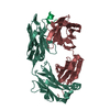
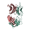
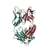
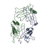
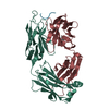
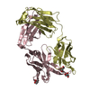
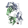
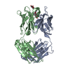
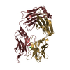
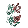
- PDB-1gpo: CRYSTAL STRUCTURE OF THE RATIONALLY DESIGNED ANTIBODY M41 AS A FA... -
+ Open data

- Basic information
| Entry | Database: PDB / ID: 1gpo | ||||||
|---|---|---|---|---|---|---|---|
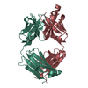
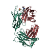
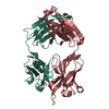
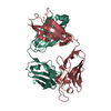
| Title | CRYSTAL STRUCTURE OF THE RATIONALLY DESIGNED ANTIBODY M41 AS A FAB FRAGMENT | ||||||
 Components Components | (ANTIBODY M41) x 2 | ||||||
 Keywords Keywords | IMMUNE SYSTEM / PROTEIN ENGINEERING / ANTIBODY DESIGN / IMMUNOGLOBULIN STRUCTURE / ANTIGEN-BINDING SITE / CANONICAL CONFORMATION / COMPLEMENTARITY-DETERMINING REGION | ||||||
| Function / homology |  Function and homology information Function and homology informationalpha-beta T cell receptor complex / IgG immunoglobulin complex / B cell differentiation / adaptive immune response / extracellular region / plasma membrane Similarity search - Function | ||||||
| Biological species |  | ||||||
| Method |  X-RAY DIFFRACTION / MOLECULAR REPLACEMENT / Resolution: 1.95 Å X-RAY DIFFRACTION / MOLECULAR REPLACEMENT / Resolution: 1.95 Å | ||||||
 Authors Authors | Schiweck, W. / Skerra, A. | ||||||
 Citation Citation | Journal: J.Mol.Biol. / Year: 1997 Title: The rational construction of an antibody against cystatin: lessons from the crystal structure of an artificial Fab fragment. Authors: Schiweck, W. / Skerra, A. #1: Journal: Proteins / Year: 1995Title: Fermenter Production of an Artificial Fab Fragment, Rationally Designed for the Antigen Cystatin, and its Optimized Crystallization Through Constant Domain Shuffling Authors: Schiweck, W. / Skerra, A. | ||||||
| History |
|
- Structure visualization
| Structure viewer | Molecule: MolmilJmol/JSmol |
|---|
- Downloads & links
-Download
| PDBx/mmCIF format | 1gpo.cif.gz | 192 KB | Display | PDBx/mmCIF format |
|---|---|---|---|---|
| PDB format | pdb1gpo.ent.gz | 151.3 KB | Display | PDB format |
| PDBx/mmJSON format | 1gpo.json.gz | Tree view | PDBx/mmJSON format | |
| Others |  Other downloads Other downloads |
-Validation report
| Arichive directory | https://data.pdbj.org/pub/pdb/validation_reports/gp/1gpoftp://data.pdbj.org/pub/pdb/validation_reports/gp/1gpo | HTTPS FTP |
|---|
-Related structure data
| Similar structure data |
|---|


-Links
 PDBj
PDBj


- Assembly
| Deposited unit | 
| ||||||||
|---|---|---|---|---|---|---|---|---|---|
| 1 | 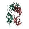
| ||||||||
| 2 | 
| ||||||||
| Unit cell |
| ||||||||
| Noncrystallographic symmetry (NCS) | NCS oper: (Code: given Matrix: (-0.997725, 0.020407, -0.064249), Vector: |
-Components
| #1: Antibody | Mass: 24172.518 Da / Num. of mol.: 2 / Fragment: FAB FRAGMENT Source method: isolated from a genetically manipulated source Source: (gene. exp.)  #2: Antibody | Mass: 24182.809 Da / Num. of mol.: 2 / Fragment: FAB FRAGMENT Source method: isolated from a genetically manipulated source Source: (gene. exp.) #3: Chemical |   Mass: 96.063 Da / Num. of mol.: 2 / Source method: obtained synthetically / Formula: SO4 Mass: 96.063 Da / Num. of mol.: 2 / Source method: obtained synthetically / Formula: SO4#4: Water | ChemComp-HOH / |  Mass: 18.015 Da / Num. of mol.: 649 / Source method: isolated from a natural source / Formula: H2O Mass: 18.015 Da / Num. of mol.: 649 / Source method: isolated from a natural source / Formula: H2OHas protein modification | Y | |
|---|
-Experimental details
-Experiment
| Experiment | Method: X-RAY DIFFRACTION / Number of used crystals: 1 |
|---|
- Sample preparation
| Crystal | Density Matthews: 2.94 Å3/Da / Density % sol: 58 % | ||||||||||||||||||||
|---|---|---|---|---|---|---|---|---|---|---|---|---|---|---|---|---|---|---|---|---|---|
| Crystal grow | pH: 6.5 Details: 2.0 M AMMONIUM SULFATE, 5 % DMSO, 0.1 M CACODYLATE PH 6.5 | ||||||||||||||||||||
| Crystal | *PLUS | ||||||||||||||||||||
| Crystal grow | *PLUS Temperature: 18 ℃ / Method: vapor diffusion, sitting dropDetails: Schiweck, W., (1995) Proteins: Struct.,Funct., Genet., 23, 561. | ||||||||||||||||||||
| Components of the solutions | *PLUS
|
-Data collection
| Diffraction | Mean temperature: 100 K |
|---|---|
| Diffraction source | Source: ROTATING ANODE / Type: RIGAKU RUH2R / Wavelength: 1.5418 |
| Detector | Type: RIGAKU / Detector: IMAGE PLATE / Date: Mar 1, 1995 |
| Radiation | Monochromator: NICKEL FILTER / Monochromatic (M) / Laue (L): M / Scattering type: x-ray |
| Radiation wavelength | Wavelength: 1.5418 Å / Relative weight: 1 |
| Reflection | Resolution: 1.95→60 Å / Num. obs: 69136 / % possible obs: 84.6 % / Observed criterion σ(I): 0.5 / Redundancy: 2.6 % / Rmerge(I) obs: 0.065 |
| Reflection shell | Resolution: 1.95→2 Å / Redundancy: 1.5 % / Rmerge(I) obs: 0.215 / % possible all: 50.2 |
- Processing
| Software |
| ||||||||||||||||||||||||||||||||||||||||||||||||||||||||||||
|---|---|---|---|---|---|---|---|---|---|---|---|---|---|---|---|---|---|---|---|---|---|---|---|---|---|---|---|---|---|---|---|---|---|---|---|---|---|---|---|---|---|---|---|---|---|---|---|---|---|---|---|---|---|---|---|---|---|---|---|---|---|
| Refinement | Method to determine structure: MOLECULAR REPLACEMENT Starting model: HYBRID FAB FRAGMENT CONSISTING OF THE MODELLED STRUCTURE FOR THE ARTIFICIAL FV FRAGMENT M41 AND CONSTANT DOMAINS OF THE ANTI-LYSOZYME ANTIBODY HYHEL-10 Resolution: 1.95→8 Å / σ(F): 2
| ||||||||||||||||||||||||||||||||||||||||||||||||||||||||||||
| Displacement parameters | Biso mean: 28.8 Å2 | ||||||||||||||||||||||||||||||||||||||||||||||||||||||||||||
| Refine analyze |
| ||||||||||||||||||||||||||||||||||||||||||||||||||||||||||||
| Refinement step | Cycle: LAST / Resolution: 1.95→8 Å
| ||||||||||||||||||||||||||||||||||||||||||||||||||||||||||||
| Refine LS restraints |
| ||||||||||||||||||||||||||||||||||||||||||||||||||||||||||||
| Software | *PLUS Name: X-PLOR / Version: 3.1 / Classification: refinement | ||||||||||||||||||||||||||||||||||||||||||||||||||||||||||||
| Refine LS restraints | *PLUS
|
