 Movie
Movie Controller

[English] 日本語
 Yorodumi
- PDB-1fux: CRYSTAL STRUCTURE OF E.COLI YBCL, A NEW MEMBER OF THE MAMMALIAN P... -
+ Open data

- Basic information
| Entry | Database: PDB / ID: 1fux | ||||||
|---|---|---|---|---|---|---|---|
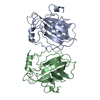
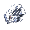
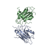
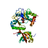
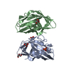
| Title | CRYSTAL STRUCTURE OF E.COLI YBCL, A NEW MEMBER OF THE MAMMALIAN PEBP FAMILY | ||||||
 Components Components | HYPOTHETICAL 19.5 KDA PROTEIN IN EMRE-RUS INTERGENIC REGION | ||||||
 Keywords Keywords | UNKNOWN FUNCTION / BETA PROTEIN | ||||||
| Function / homology |  Function and homology information Function and homology information | ||||||
| Biological species |  | ||||||
| Method |  X-RAY DIFFRACTION / SYNCHROTRON / MOLECULAR REPLACEMENT / Resolution: 1.81 Å X-RAY DIFFRACTION / SYNCHROTRON / MOLECULAR REPLACEMENT / Resolution: 1.81 Å | ||||||
 Authors Authors | Serre, L. / Pereira de Jesus, K. / Benedetti, H. / Bureaud, N. / Schoentgen, F. / Zelwer, C. | ||||||
 Citation Citation | Journal: J.Mol.Biol. / Year: 2001 Title: Crystal structures of YBHB and YBCL from Escherichia coli, two bacterial homologues to a Raf kinase inhibitor protein. Authors: Serre, L. / Pereira de Jesus, K. / Zelwer, C. / Bureaud, N. / Schoentgen, F. / Benedetti, H. | ||||||
| History |
|
- Structure visualization
| Structure viewer | Molecule: MolmilJmol/JSmol |
|---|
- Downloads & links
-Download
| PDBx/mmCIF format | 1fux.cif.gz | 79.2 KB | Display | PDBx/mmCIF format |
|---|---|---|---|---|
| PDB format | pdb1fux.ent.gz | 58.7 KB | Display | PDB format |
| PDBx/mmJSON format | 1fux.json.gz | Tree view | PDBx/mmJSON format | |
| Others |  Other downloads Other downloads |
-Validation report
| Arichive directory | https://data.pdbj.org/pub/pdb/validation_reports/fu/1fuxftp://data.pdbj.org/pub/pdb/validation_reports/fu/1fux | HTTPS FTP |
|---|
-Related structure data
| Related structure data |  1fjjSC S: Starting model for refinement C: citing same article ( |
|---|---|
| Similar structure data |


-Links
 PDBj
PDBj- Assembly
| Deposited unit | 
| ||||||||
|---|---|---|---|---|---|---|---|---|---|
| 1 |
| ||||||||
| Unit cell |
| ||||||||
| Details | the biological assembly is a dimer constructed from chain A and chain B |
-Components
| #1: Protein | Mass: 17816.643 Da / Num. of mol.: 2 Source method: isolated from a genetically manipulated source Details: PERIPLASMIC FORM / Source: (gene. exp.) Keywords: PERIPLASMIC FORM / References: UniProt: P77368#2: Water | ChemComp-HOH / |  Mass: 18.015 Da / Num. of mol.: 206 / Source method: isolated from a natural source / Formula: H2O Mass: 18.015 Da / Num. of mol.: 206 / Source method: isolated from a natural source / Formula: H2OHas protein modification | Y | Nonpolymer details | MOLECULES 401, 402, 403, 404 HAVE BEEN MODELED BY HOH BUT CORRESPOND TO UNKNOWN MOLECULES PRESENT ...MOLECULES 401, 402, 403, 404 HAVE BEEN MODELED BY HOH BUT CORRESPOND | |
|---|
-Experimental details
-Experiment
| Experiment | Method: X-RAY DIFFRACTION / Number of used crystals: 1 |
|---|
- Sample preparation
| Crystal | Density Matthews: 2.2 Å3/Da / Density % sol: 44 % | ||||||||||||||||||||||||||||||
|---|---|---|---|---|---|---|---|---|---|---|---|---|---|---|---|---|---|---|---|---|---|---|---|---|---|---|---|---|---|---|---|
| Crystal grow | Temperature: 298 K / Method: vapor diffusion, hanging drop / pH: 7.3 Details: PEG 6000, Hepes, pH 7.3, VAPOR DIFFUSION, HANGING DROP, temperature 298.0K | ||||||||||||||||||||||||||||||
| Crystal grow | *PLUS pH: 7 | ||||||||||||||||||||||||||||||
| Components of the solutions | *PLUS
|
-Data collection
| Diffraction | Mean temperature: 100 K |
|---|---|
| Diffraction source | Source: SYNCHROTRON / Site: ESRF  / Beamline: BM30A / Wavelength: 0.97941 / Beamline: BM30A / Wavelength: 0.97941 |
| Detector | Type: MARRESEARCH / Detector: IMAGE PLATE / Date: Jun 23, 2000 |
| Radiation | Protocol: SINGLE WAVELENGTH / Monochromatic (M) / Laue (L): M / Scattering type: x-ray |
| Radiation wavelength | Wavelength: 0.97941 Å / Relative weight: 1 |
| Reflection | Resolution: 1.81→25 Å / Num. obs: 96044 / % possible obs: 97.5 % / Observed criterion σ(F): 0 / Observed criterion σ(I): 0 / Redundancy: 3.4 % / Biso Wilson estimate: 16.894 Å2 / Rsym value: 0.03 / Net I/σ(I): 16.1 |
| Reflection shell | Resolution: 1.81→1.91 Å / Redundancy: 3.2 % / Mean I/σ(I) obs: 7.8 / Num. unique all: 3751 / Rsym value: 0.088 / % possible all: 89.4 |
| Reflection | *PLUS Lowest resolution: 25 Å / Num. obs: 28148 / Num. measured all: 96044 / Rmerge(I) obs: 0.03 |
| Reflection shell | *PLUS Rmerge(I) obs: 0.088 |
- Processing
| Software |
| ||||||||||||||||||||||||||||||||||||||||||||||||||||||||||||
|---|---|---|---|---|---|---|---|---|---|---|---|---|---|---|---|---|---|---|---|---|---|---|---|---|---|---|---|---|---|---|---|---|---|---|---|---|---|---|---|---|---|---|---|---|---|---|---|---|---|---|---|---|---|---|---|---|---|---|---|---|---|
| Refinement | Method to determine structure: MOLECULAR REPLACEMENT Starting model: 1FJJ E.COLI YBHB Resolution: 1.81→25 Å / SU B: 3.31573 / SU ML: 0.10434 / Cross valid method: FREE-R / σ(F): 0 / σ(I): 0 / ESU R: 0.16318 / ESU R Free: 0.15721 / Stereochemistry target values: Engh & Huber
| ||||||||||||||||||||||||||||||||||||||||||||||||||||||||||||
| Displacement parameters | Biso mean: 21.61 Å2 | ||||||||||||||||||||||||||||||||||||||||||||||||||||||||||||
| Refinement step | Cycle: LAST / Resolution: 1.81→25 Å
| ||||||||||||||||||||||||||||||||||||||||||||||||||||||||||||
| Refine LS restraints |
| ||||||||||||||||||||||||||||||||||||||||||||||||||||||||||||
| Software | *PLUS Name: REFMAC / Classification: refinement | ||||||||||||||||||||||||||||||||||||||||||||||||||||||||||||
| Refinement | *PLUS Rfactor obs: 0.199 | ||||||||||||||||||||||||||||||||||||||||||||||||||||||||||||
| Solvent computation | *PLUS | ||||||||||||||||||||||||||||||||||||||||||||||||||||||||||||
| Displacement parameters | *PLUS | ||||||||||||||||||||||||||||||||||||||||||||||||||||||||||||
| Refine LS restraints | *PLUS
|
