 Movie
Movie Controller


+ Open data

- Basic information
| Entry | Database: PDB / ID: 1qou | ||||||
|---|---|---|---|---|---|---|---|
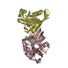
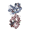
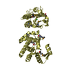
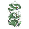
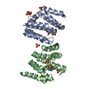
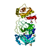
| Title | CEN (Centroradialis) protein from Antirrhinum | ||||||
 Components Components | CEN | ||||||
 Keywords Keywords | PLANT PROTEIN / INFLUORESCENCE DETERMINATION IN FLOWERING PLANT MERISTEM / SIGNALLING / MEMBER OF THE PHOSPHATIDYLETHANOLAMINE BINDING PROTEIN FAMILY | ||||||
| Function / homology |  Function and homology information Function and homology informationnegative regulation of flower development / vegetative to reproductive phase transition of meristem / flower development / cell differentiation / nucleus / cytoplasm Similarity search - Function | ||||||
| Biological species |  ANTIRRHINUM MAJUS (snapdragon) ANTIRRHINUM MAJUS (snapdragon) | ||||||
| Method |  X-RAY DIFFRACTION / SYNCHROTRON / MOLECULAR REPLACEMENT / Resolution: 1.9 Å X-RAY DIFFRACTION / SYNCHROTRON / MOLECULAR REPLACEMENT / Resolution: 1.9 Å | ||||||
 Authors Authors | Banfield, M.J. / Brady, R.L. | ||||||
 Citation Citation | Journal: J.Mol.Biol. / Year: 2000 Title: The Structure of Antirrhinum Centroradialis Protein (Cen) Suggests a Role as a Kinase Regulator Authors: Banfield, M.J. / Brady, R.L. #1: Journal: Structure / Year: 1998Title: Function from Structure? the Crystal Structure of Human Phosphatidylethanolamine Binding Protein Suggests a Role in Membrane Signal Transduction Authors: Banfield, M.J. / Barker, J.J. / Perry, A. / Brady, R.L. #2: Journal: Structure / Year: 1998Title: Crystal Structure of Bovine Phosphatidylethanolamine-Binding Proteins from Bovine Brain: A Novel Structural Class of Phospholipid-Binding Proteins. Authors: Serre, L. / Vallee, B. / Bureaud, N. / Schoentgen, F. / Zelwer, C. | ||||||
| History |
|
- Structure visualization
| Structure viewer | Molecule: MolmilJmol/JSmol |
|---|
- Downloads & links
-Download
| PDBx/mmCIF format | 1qou.cif.gz | 80.4 KB | Display | PDBx/mmCIF format |
|---|---|---|---|---|
| PDB format | pdb1qou.ent.gz | 59.7 KB | Display | PDB format |
| PDBx/mmJSON format | 1qou.json.gz | Tree view | PDBx/mmJSON format | |
| Others |  Other downloads Other downloads |
-Validation report
| Arichive directory | https://data.pdbj.org/pub/pdb/validation_reports/qo/1qouftp://data.pdbj.org/pub/pdb/validation_reports/qo/1qou | HTTPS FTP |
|---|
-Related structure data
| Related structure data | 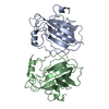 1bd9S S: Starting model for refinement |
|---|---|
| Similar structure data |


-Links
 PDBj
PDBj- Assembly
| Deposited unit | 
| ||||||||
|---|---|---|---|---|---|---|---|---|---|
| 1 |
| ||||||||
| Unit cell |
| ||||||||
| Noncrystallographic symmetry (NCS) | NCS oper: (Code: given Matrix: (-0.997832, 0.065499, 0.006386), Vector: Details | IN THE CRYSTAL THE PROTEIN FORMS A HOMO- DIMER THROUGHAN INTER-CHAIN DISULPHIDE BOND. THIS OLIGOMERIC STATEIS NOT EXPECTED TO REPRESENT THE FUNCTIONAL STATE.HOWEVER, A ROLE FOR THIS DIMERIC ASSOCIATION IN PROTEINFUNCTION CANNOT BE RULED OUT . | |
-Components
| #1: Protein | Mass: 20350.279 Da / Num. of mol.: 2 Source method: isolated from a genetically manipulated source Details: INTER-MOLECULAR DISULPHIDE BOND FORMED IN THE CRYSTAL, BETWEEN RESIDUES A145 AND B145. Source: (gene. exp.) ANTIRRHINUM MAJUS (snapdragon) / Cellular location: CYTOPLASM / Cellular location (production host): CYTOPLASM / Production host:  #2: Water | ChemComp-HOH / |  Mass: 18.015 Da / Num. of mol.: 283 / Source method: isolated from a natural source / Formula: H2O Mass: 18.015 Da / Num. of mol.: 283 / Source method: isolated from a natural source / Formula: H2OHas protein modification | Y | Sequence details | AMINO ACID NUMBERING: FOR EASE OF COMPARISON, THE NUMBERING SCHEME ADOPTED FOR THIS ENTRY IS BASED ...AMINO ACID NUMBERING: FOR EASE OF COMPARISON | |
|---|
-Experimental details
-Experiment
| Experiment | Method: X-RAY DIFFRACTION / Number of used crystals: 1 |
|---|
- Sample preparation
| Crystal | Density Matthews: 2.3 Å3/Da / Density % sol: 39.3 % | ||||||||||||||||||||||||||||||||||||
|---|---|---|---|---|---|---|---|---|---|---|---|---|---|---|---|---|---|---|---|---|---|---|---|---|---|---|---|---|---|---|---|---|---|---|---|---|---|
| Crystal grow | pH: 5.5 Details: 1:1 MIX OF 13MG/ML CEN SOLUTION (10MM HEPES, 50MM NACL. PH 7) WITH 1.8 - 2.1 M NACL BUFFERED WITH 100MM SODIUM ACETATE AT PH 5.4 - 5.6 | ||||||||||||||||||||||||||||||||||||
| Crystal | *PLUS | ||||||||||||||||||||||||||||||||||||
| Crystal grow | *PLUS pH: 7 / Method: unknown | ||||||||||||||||||||||||||||||||||||
| Components of the solutions | *PLUS
|
-Data collection
| Diffraction | Mean temperature: 100 K |
|---|---|
| Diffraction source | Source: SYNCHROTRON / Site: SRS  / Beamline: PX7.2 / Wavelength: 1.488 / Beamline: PX7.2 / Wavelength: 1.488 |
| Detector | Type: MARRESEARCH / Detector: IMAGE PLATE / Date: Mar 15, 1999 / Details: MIRRORS |
| Radiation | Protocol: SINGLE WAVELENGTH / Monochromatic (M) / Laue (L): M / Scattering type: x-ray |
| Radiation wavelength | Wavelength: 1.488 Å / Relative weight: 1 |
| Reflection | Resolution: 1.9→40 Å / Num. obs: 28860 / % possible obs: 94.9 % / Redundancy: 4.2 % / Biso Wilson estimate: 19.29 Å2 / Rmerge(I) obs: 0.057 / Net I/σ(I): 22.4 |
| Reflection shell | Resolution: 1.9→1.99 Å / Redundancy: 3.5 % / Rmerge(I) obs: 0.226 / Mean I/σ(I) obs: 4.6 / % possible all: 91.4 |
| Reflection shell | *PLUS % possible obs: 91.4 % |
- Processing
| Software |
| ||||||||||||||||||||||||||||||||||||||||||||||||||||||||||||||||||||||||||||||||||||
|---|---|---|---|---|---|---|---|---|---|---|---|---|---|---|---|---|---|---|---|---|---|---|---|---|---|---|---|---|---|---|---|---|---|---|---|---|---|---|---|---|---|---|---|---|---|---|---|---|---|---|---|---|---|---|---|---|---|---|---|---|---|---|---|---|---|---|---|---|---|---|---|---|---|---|---|---|---|---|---|---|---|---|---|---|---|
| Refinement | Method to determine structure: MOLECULAR REPLACEMENT Starting model: 1BD9 Resolution: 1.9→40 Å / SU B: 3.65 / SU ML: 0.11 / Cross valid method: THROUGHOUT / σ(F): 0 / ESU R: 0.17 / ESU R Free: 0.16 Details: THE REFINED MODEL OF THE A-CHAIN CONTAINS THE FOLLOWING RESIDUES OF THE SEQUENCE LISTED REFINED MODEL OF THE B-CHAIN CONTAINS THE FOLLOWING RESIDUES B32B- B130, B143-B175.
| ||||||||||||||||||||||||||||||||||||||||||||||||||||||||||||||||||||||||||||||||||||
| Displacement parameters | Biso mean: 27.44 Å2 | ||||||||||||||||||||||||||||||||||||||||||||||||||||||||||||||||||||||||||||||||||||
| Refinement step | Cycle: LAST / Resolution: 1.9→40 Å
| ||||||||||||||||||||||||||||||||||||||||||||||||||||||||||||||||||||||||||||||||||||
| Refine LS restraints |
| ||||||||||||||||||||||||||||||||||||||||||||||||||||||||||||||||||||||||||||||||||||
| Software | *PLUS Name: REFMAC / Classification: refinement | ||||||||||||||||||||||||||||||||||||||||||||||||||||||||||||||||||||||||||||||||||||
| Refinement | *PLUS Rfactor obs: 0.212 / Rfactor Rfree: 0.25 | ||||||||||||||||||||||||||||||||||||||||||||||||||||||||||||||||||||||||||||||||||||
| Solvent computation | *PLUS | ||||||||||||||||||||||||||||||||||||||||||||||||||||||||||||||||||||||||||||||||||||
| Displacement parameters | *PLUS | ||||||||||||||||||||||||||||||||||||||||||||||||||||||||||||||||||||||||||||||||||||
| LS refinement shell | *PLUS Rfactor Rfree: 0.303 / Rfactor obs: 0.259 |
