 Movie
Movie Controller

[English] 日本語
 Yorodumi
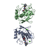
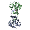
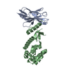
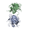
- PDB-1b7a: STRUCTURE OF THE PHOSPHATIDYLETHANOLAMINE-BINDING PROTEIN FROM BO... -
+ Open data

- Basic information
| Entry | Database: PDB / ID: 1b7a | ||||||
|---|---|---|---|---|---|---|---|
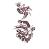
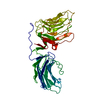
| Title | STRUCTURE OF THE PHOSPHATIDYLETHANOLAMINE-BINDING PROTEIN FROM BOVINE BRAIN | ||||||
 Components Components | PHOSPHATIDYLETHANOLAMINE-BINDING PROTEIN | ||||||
 Keywords Keywords | LIPID BINDING PROTEIN / PHOSPHOLIPID BINDING PROTEIN | ||||||
| Function / homology |  Function and homology information Function and homology informationMAP2K and MAPK activation / Negative regulation of MAPK pathway / negative regulation of MAPK cascade / serine-type endopeptidase inhibitor activity / lipid binding / ATP binding / cytoplasm Similarity search - Function | ||||||
| Biological species |  | ||||||
| Method |  X-RAY DIFFRACTION / SYNCHROTRON / MOLECULAR REPLACEMENT / Resolution: 2.25 Å X-RAY DIFFRACTION / SYNCHROTRON / MOLECULAR REPLACEMENT / Resolution: 2.25 Å | ||||||
 Authors Authors | Serre, L. / Vallee, B. / Bureaud, N. / Schoentgen, F. / Zelwer, C. | ||||||
 Citation Citation | Journal: Structure / Year: 1998 Title: Crystal structure of the phosphatidylethanolamine-binding protein from bovine brain: a novel structural class of phospholipid-binding proteins. Authors: Serre, L. / Vallee, B. / Bureaud, N. / Schoentgen, F. / Zelwer, C. | ||||||
| History |
|
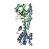
- Structure visualization
| Structure viewer | Molecule: MolmilJmol/JSmol |
|---|
- Downloads & links
-Download
| PDBx/mmCIF format | 1b7a.cif.gz | 89.1 KB | Display | PDBx/mmCIF format |
|---|---|---|---|---|
| PDB format | pdb1b7a.ent.gz | 67.3 KB | Display | PDB format |
| PDBx/mmJSON format | 1b7a.json.gz | Tree view | PDBx/mmJSON format | |
| Others |  Other downloads Other downloads |
-Validation report
| Arichive directory | https://data.pdbj.org/pub/pdb/validation_reports/b7/1b7aftp://data.pdbj.org/pub/pdb/validation_reports/b7/1b7a | HTTPS FTP |
|---|
-Related structure data
| Related structure data |  1a44SC S: Starting model for refinement C: citing same article ( |
|---|---|
| Similar structure data |


-Links
 PDBj
PDBj
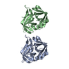
- Assembly
| Deposited unit | 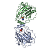
| ||||||||||
|---|---|---|---|---|---|---|---|---|---|---|---|
| 1 |
| ||||||||||
| Unit cell |
| ||||||||||
| Noncrystallographic symmetry (NCS) | NCS oper: (Code: given Matrix: (-0.97096, -0.23875, -0.01506), Vector: |
-Components
| #1: Protein | Mass: 20883.543 Da / Num. of mol.: 2 / Source method: isolated from a natural source / Source: (natural) #2: Chemical | 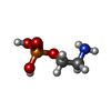  Mass: 141.063 Da / Num. of mol.: 2 / Source method: obtained synthetically / Formula: C2H8NO4P Mass: 141.063 Da / Num. of mol.: 2 / Source method: obtained synthetically / Formula: C2H8NO4P#3: Water | ChemComp-HOH / |  Mass: 18.015 Da / Num. of mol.: 164 / Source method: isolated from a natural source / Formula: H2O Mass: 18.015 Da / Num. of mol.: 164 / Source method: isolated from a natural source / Formula: H2ONonpolymer details | THE OPE MOLECULES HAVE TWO CONFORMATI | Sequence details | GLY: THE SIDE CHAIN OF THE C-TERMINAL LYS186 WAS NOT SEEN I THE ELECTRON DENSITY MAP AND WAS ...GLY: THE SIDE CHAIN OF THE C-TERMINAL LYS186 WAS NOT SEEN I THE ELECTRON DENSITY MAP AND WAS REFINED AS GLYCINE. | |
|---|
-Experimental details
-Experiment
| Experiment | Method: X-RAY DIFFRACTION / Number of used crystals: 1 |
|---|
- Sample preparation
| Crystal | Density Matthews: 2.18 Å3/Da / Density % sol: 44 % | ||||||||||||||||||||||||||||||
|---|---|---|---|---|---|---|---|---|---|---|---|---|---|---|---|---|---|---|---|---|---|---|---|---|---|---|---|---|---|---|---|
| Crystal grow | pH: 4.3 Details: 25-27% PEG8000, 100 MM PHOSPHORYLETHANOLAMINE, pH 4.3 | ||||||||||||||||||||||||||||||
| Components of the solutions |
| ||||||||||||||||||||||||||||||
| Crystal grow | *PLUS pH: 7 / Method: vapor diffusion, hanging drop | ||||||||||||||||||||||||||||||
| Components of the solutions | *PLUS
|
-Data collection
| Diffraction | Mean temperature: 100 K |
|---|---|
| Diffraction source | Source: SYNCHROTRON / Site: LURE  / Beamline: DW32 / Wavelength: 1 / Beamline: DW32 / Wavelength: 1 |
| Detector | Type: MARRESEARCH / Detector: IMAGE PLATE / Date: May 15, 1998 |
| Radiation | Protocol: SINGLE WAVELENGTH / Monochromatic (M) / Laue (L): M / Scattering type: x-ray |
| Radiation wavelength | Wavelength: 1 Å / Relative weight: 1 |
| Reflection | Resolution: 2.25→29 Å / Num. obs: 16450 / % possible obs: 90.8 % / Observed criterion σ(I): 1 / Redundancy: 3.9 % / Biso Wilson estimate: 24.7 Å2 / Rmerge(I) obs: 0.057 / Rsym value: 4.9 / Net I/σ(I): 12.4 |
| Reflection shell | Resolution: 2.25→2.41 Å / Redundancy: 3.8 % / Rmerge(I) obs: 0.122 / Mean I/σ(I) obs: 7.1 / Rsym value: 10.6 / % possible all: 88.8 |
- Processing
| Software |
| ||||||||||||||||||||||||||||||||||||||||||||||||||||||||||||||||||||||||||||||||||||
|---|---|---|---|---|---|---|---|---|---|---|---|---|---|---|---|---|---|---|---|---|---|---|---|---|---|---|---|---|---|---|---|---|---|---|---|---|---|---|---|---|---|---|---|---|---|---|---|---|---|---|---|---|---|---|---|---|---|---|---|---|---|---|---|---|---|---|---|---|---|---|---|---|---|---|---|---|---|---|---|---|---|---|---|---|---|
| Refinement | Method to determine structure: MOLECULAR REPLACEMENT Starting model: PDB ENTRY 1A44 Resolution: 2.25→12.5 Å / Cross valid method: THROUGHOUT / σ(F): 0
| ||||||||||||||||||||||||||||||||||||||||||||||||||||||||||||||||||||||||||||||||||||
| Displacement parameters | Biso mean: 24.37 Å2 | ||||||||||||||||||||||||||||||||||||||||||||||||||||||||||||||||||||||||||||||||||||
| Refinement step | Cycle: LAST / Resolution: 2.25→12.5 Å
| ||||||||||||||||||||||||||||||||||||||||||||||||||||||||||||||||||||||||||||||||||||
| Refine LS restraints |
| ||||||||||||||||||||||||||||||||||||||||||||||||||||||||||||||||||||||||||||||||||||
| Software | *PLUS Name: REFMAC / Classification: refinement | ||||||||||||||||||||||||||||||||||||||||||||||||||||||||||||||||||||||||||||||||||||
| Refinement | *PLUS σ(F): 0 / % reflection Rfree: 10 % / Rfactor obs: 0.208 | ||||||||||||||||||||||||||||||||||||||||||||||||||||||||||||||||||||||||||||||||||||
| Solvent computation | *PLUS | ||||||||||||||||||||||||||||||||||||||||||||||||||||||||||||||||||||||||||||||||||||
| Displacement parameters | *PLUS |
