SEQUENCE THE CONSTRUCT WAS EXPRESSED WITH A PURIFICATION TAG MGSDKIHHHHHHENLYFQG. THE TAG WAS ...SEQUENCE THE CONSTRUCT WAS EXPRESSED WITH A PURIFICATION TAG MGSDKIHHHHHHENLYFQG. THE TAG WAS REMOVED WITH TEV PROTEASE LEAVING ONLY A GLYCINE (0) FOLLOWED BY THE TARGET SEQUENCE. THE STRAIN CLONED DIFFERS FROM THE SEQUENCED STRAIN IN THE DATABASE. THE ELECTRON DENSITY CLEARLY INDICATED THAT VALINE AT 45 SHOULD BE ISOLEUCINE AND SERINE AT 102 SHOULD BE TYROSINE, I.E., V45I AND S102Y. THE DNA SEQUENCE OF THE CLONED CONSTRUCT CONFIRMS THIS OBSERVATION.
Resolution: 1.41→18.62 Å / Num. obs: 46247 / % possible obs: 83.4 % / Redundancy: 3.1 % / Rmerge(I) obs: 0.041 / Rsym value: 0.041 / Net I/σ(I): 9
Reflection shell
Resolution (Å)
% possible obs (%)
Redundancy (%)
Rmerge(I) obs
Mean I/σ(I) obs
Num. measured obs
Rsym value
% possible all
1.41-1.45
32.3
1.6
0.276
2.5
1312
0.276
32.3
1.45-1.49
41.6
1.8
0.221
3.3
1635
0.221
1.49-1.53
54.4
2
0.176
4.1
2106
0.176
1.53-1.58
66.1
2.2
0.139
5.2
2476
0.139
1.58-1.63
79.2
2.4
0.121
5.9
2839
0.121
1.63-1.69
94.5
2.8
0.106
6.5
3325
0.106
1.69-1.75
99.7
3.1
0.087
7.7
3399
0.087
1.75-1.82
99.9
3.6
0.077
7.8
3242
0.077
1.82-1.9
99.8
3.6
0.068
8.5
3151
0.068
1.9-1.99
100
3.6
0.065
9
3012
0.065
1.99-2.1
99.9
3.6
0.058
9.3
2844
0.058
2.1-2.23
100
3.7
0.048
10.7
2721
0.048
2.23-2.38
100
3.6
0.05
10.1
2560
0.05
2.38-2.57
100
3.6
0.04
12.2
2388
0.04
2.57-2.82
100
3.6
0.039
12.5
2185
0.039
2.82-3.15
100
3.6
0.035
13.5
1976
0.035
3.15-3.64
99.9
3.5
0.033
15.2
1773
0.033
3.64-4.46
99.4
3.4
0.031
16.3
1496
0.031
4.46-6.31
99.8
3.3
0.028
16.4
1177
0.028
6.31-18.62
92.4
3
0.03
15.4
630
0.03
-
Phasing
Phasing
Method: MAD
-
Processing
Software
Name
Version
Classification
NB
REFMAC
5.2.0005
refinement
SCALA
datascaling
PDB_EXTRACT
1.601
dataextraction
MOSFLM
datareduction
CCP4
(SCALA)
datascaling
SOLVE
phasing
Refinement
Method to determine structure: MAD / Resolution: 1.41→18.62 Å / Cor.coef. Fo:Fc: 0.97 / Cor.coef. Fo:Fc free: 0.963 / SU B: 2.719 / SU ML: 0.054 / Cross valid method: THROUGHOUT / ESU R: 0.09 / ESU R Free: 0.089 Stereochemistry target values: MAXIMUM LIKELIHOOD WITH PHASES Details: 1. HYDROGENS HAVE BEEN ADDED IN THE RIDING POSITIONS 2. THERE EXISTS A PSEUDO-TRANSLATION BETWEEN THE TWO MONOMERS IN THE ASU. AS A RESULT, THE L=2N+1 REFLECTIONS ARE SYSTEMATICALLY WEAK. ...Details: 1. HYDROGENS HAVE BEEN ADDED IN THE RIDING POSITIONS 2. THERE EXISTS A PSEUDO-TRANSLATION BETWEEN THE TWO MONOMERS IN THE ASU. AS A RESULT, THE L=2N+1 REFLECTIONS ARE SYSTEMATICALLY WEAK. THIS RESULTS IN HIGH R-FACTOR FOR L=2N+1 REFLECTIONS. THE OVERALL R-FACTORS IS RELATIVELY HIGH DUE TO THIS REASON. THE MAPS LOOK VERY GOOD. 3. AN UNKNOWN DENSITY NEAR B88 WAS MODELED AS UNL, UNKNOWN LIGAND. 4. DATA AT HIGHEST RESOLUTION SHELLS ARE INCOMPLETE.
Rfactor
Num. reflection
% reflection
Selection details
Rfree
0.228
2352
5.1 %
RANDOM
Rwork
0.2
-
-
-
all
0.201
-
-
-
obs
0.20104
43885
83.43 %
-
Solvent computation
Ion probe radii: 0.8 Å / Shrinkage radii: 0.8 Å / VDW probe radii: 1.2 Å / Solvent model: BABINET MODEL WITH MASK
In the structure databanks used in Yorodumi, some data are registered as the other names, "COVID-19 virus" and "2019-nCoV". Here are the details of the virus and the list of structure data.
Jan 31, 2019. EMDB accession codes are about to change! (news from PDBe EMDB page)
EMDB accession codes are about to change! (news from PDBe EMDB page)
The allocation of 4 digits for EMDB accession codes will soon come to an end. Whilst these codes will remain in use, new EMDB accession codes will include an additional digit and will expand incrementally as the available range of codes is exhausted. The current 4-digit format prefixed with “EMD-” (i.e. EMD-XXXX) will advance to a 5-digit format (i.e. EMD-XXXXX), and so on. It is currently estimated that the 4-digit codes will be depleted around Spring 2019, at which point the 5-digit format will come into force.
The EM Navigator/Yorodumi systems omit the EMD- prefix.
Related info.:Q: What is EMD? / ID/Accession-code notation in Yorodumi/EM Navigator
Yorodumi is a browser for structure data from EMDB, PDB, SASBDB, etc.
This page is also the successor to EM Navigator detail page, and also detail information page/front-end page for Omokage search.
The word "yorodu" (or yorozu) is an old Japanese word meaning "ten thousand". "mi" (miru) is to see.
Related info.:EMDB / PDB / SASBDB / Comparison of 3 databanks / Yorodumi Search / Aug 31, 2016. New EM Navigator & Yorodumi / Yorodumi Papers / Jmol/JSmol / Function and homology information / Changes in new EM Navigator and Yorodumi
 Movie
Movie Controller

 Yorodumi
 Open data
 Basic information
 Components
 Keywords
 Function and homology information
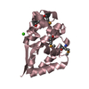
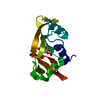
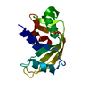
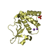
 Neisseria meningitidis Z2491 (bacteria)
 X-RAY DIFFRACTION /
  Authors
 Citation
 Structure visualization
 Downloads & links
 Other downloads
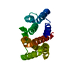


 PDBj
PDBj


 Assembly


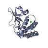
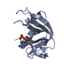
 Mass: 35.453 Da / Num. of mol.: 2 / Source method: obtained synthetically / Formula: Cl
 Mass: 18.015 Da / Num. of mol.: 409 / Source method: isolated from a natural source / Formula: H2O
 Sample preparation
 / Beamline: 8.3.1 / Wavelength: 1.019951, 0.979741
 Processing