 Movie
Movie Controller

[English] 日本語
 Yorodumi
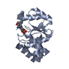
- PDB-2i6p: Crystal structure of the complex of the archaeal sulfolobus PTP-f... -
+ Open data

- Basic information
| Entry | Database: PDB / ID: 2i6p | ||||||
|---|---|---|---|---|---|---|---|
| Title | Crystal structure of the complex of the archaeal sulfolobus PTP-fold phosphatase with pNPP | ||||||
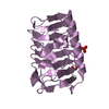
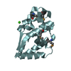
 Components Components | Sulfolobus solfataricus protein tyrosine phosphatase | ||||||
 Keywords Keywords | HYDROLASE / PTP domain / tyrosine phosphatase | ||||||
| Function / homology |  Function and homology information Function and homology information | ||||||
| Biological species |   Sulfolobus solfataricus (archaea) Sulfolobus solfataricus (archaea) | ||||||
| Method |  X-RAY DIFFRACTION / MOLECULAR REPLACEMENT / Resolution: 2.5 Å X-RAY DIFFRACTION / MOLECULAR REPLACEMENT / Resolution: 2.5 Å | ||||||
 Authors Authors | Chu, H.M. / Wang, A.H.J. | ||||||
 Citation Citation | Journal: Proteins / Year: 2006 Title: Enzyme-substrate interactions revealed by the crystal structures of the archaeal Sulfolobus PTP-fold phosphatase and its phosphopeptide complexes Authors: Chu, H.M. / Wang, A.H.J. | ||||||
| History |
|
- Structure visualization
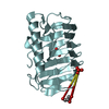
| Structure viewer | Molecule: MolmilJmol/JSmol |
|---|
- Downloads & links
-Download
| PDBx/mmCIF format | 2i6p.cif.gz | 45.9 KB | Display | PDBx/mmCIF format |
|---|---|---|---|---|
| PDB format | pdb2i6p.ent.gz | 31.5 KB | Display | PDB format |
| PDBx/mmJSON format | 2i6p.json.gz | Tree view | PDBx/mmJSON format | |
| Others |  Other downloads Other downloads |
-Validation report
| Arichive directory | https://data.pdbj.org/pub/pdb/validation_reports/i6/2i6pftp://data.pdbj.org/pub/pdb/validation_reports/i6/2i6p | HTTPS FTP |
|---|
-Related structure data
| Related structure data |  2dxpC 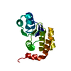 2i6iC 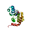 2i6jC 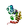 2i6mC 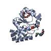 2i6oC  1oheS C: citing same article ( S: Starting model for refinement |
|---|---|
| Similar structure data |


-Links
 PDBj
PDBj


- Assembly
| Deposited unit | 
| ||||||||
|---|---|---|---|---|---|---|---|---|---|
| 1 |
| ||||||||
| Unit cell |
|
-Components
| #1: Protein | Mass: 18417.100 Da / Num. of mol.: 1 / Mutation: C96S Source method: isolated from a genetically manipulated source Source: (gene. exp.) Sulfolobus solfataricus (archaea) / Plasmid: pET21 / Species (production host): Escherichia coli / Production host:  |
|---|---|
| #2: Chemical | ChemComp-4NP / 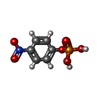  Mass: 219.089 Da / Num. of mol.: 1 / Source method: obtained synthetically / Formula: C6H6NO6P Mass: 219.089 Da / Num. of mol.: 1 / Source method: obtained synthetically / Formula: C6H6NO6P |
| #3: Water | ChemComp-HOH /  Mass: 18.015 Da / Num. of mol.: 32 / Source method: isolated from a natural source / Formula: H2O Mass: 18.015 Da / Num. of mol.: 32 / Source method: isolated from a natural source / Formula: H2O |
-Experimental details
-Experiment
| Experiment | Method: X-RAY DIFFRACTION / Number of used crystals: 1 |
|---|
- Sample preparation
| Crystal | Density Matthews: 2.27 Å3/Da / Density % sol: 45.87 % |
|---|---|
| Crystal grow | Temperature: 298 K / Method: vapor diffusion, hanging drop / pH: 7.5 Details: pH 7.5, VAPOR DIFFUSION, HANGING DROP, temperature 298K |
-Data collection
| Diffraction | Mean temperature: 298 K |
|---|---|
| Diffraction source | Source: ROTATING ANODE / Type: RIGAKU |
| Detector | Type: RIGAKU RAXIS IV / Detector: IMAGE PLATE / Date: Aug 1, 2006 |
| Radiation | Protocol: SINGLE WAVELENGTH / Monochromatic (M) / Laue (L): M / Scattering type: x-ray |
| Radiation wavelength | Relative weight: 1 |
| Reflection | Resolution: 2.5→50 Å / Num. all: 5786 / Num. obs: 4664 / % possible obs: 80.6 % / Observed criterion σ(F): 2 / Redundancy: 2.8 % / Rmerge(I) obs: 0.077 / Rsym value: 0.062 / Net I/σ(I): 17.6 |
| Reflection shell | Resolution: 2.5→2.59 Å / Redundancy: 2.5 % / Rmerge(I) obs: 0.319 / Mean I/σ(I) obs: 2.77 / Num. unique all: 517 / Rsym value: 0.279 / % possible all: 91.7 |
- Processing
| Software |
| ||||||||||||||||||||
|---|---|---|---|---|---|---|---|---|---|---|---|---|---|---|---|---|---|---|---|---|---|
| Refinement | Method to determine structure: MOLECULAR REPLACEMENT Starting model: 1OHE Resolution: 2.5→30 Å / σ(F): 2
| ||||||||||||||||||||
| Refinement step | Cycle: LAST / Resolution: 2.5→30 Å
| ||||||||||||||||||||
| Refine LS restraints |
|
