 Movie
Movie Controller


+ Open data

- Basic information
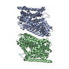
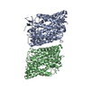
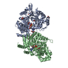
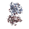
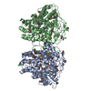
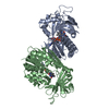
| Entry | Database: PDB / ID: 1f3j | ||||||
|---|---|---|---|---|---|---|---|
| Title | HISTOCOMPATIBILITY ANTIGEN I-AG7 | ||||||
 Components Components |
| ||||||
 Keywords Keywords | IMMUNE SYSTEM / HISTOCOMPATIBILITY ANTIGEN / MHC / PEPTIDE COMPLEX | ||||||
| Function / homology |  Function and homology information Function and homology informationantigen processing and presentation of peptide antigen / positive regulation of T cell differentiation / antigen processing and presentation / multivesicular body / Lactose synthesis / Antimicrobial peptides / Neutrophil degranulation / beta-N-acetylglucosaminidase activity / MHC class II protein complex / antigen processing and presentation of exogenous peptide antigen via MHC class II ...antigen processing and presentation of peptide antigen / positive regulation of T cell differentiation / antigen processing and presentation / multivesicular body / Lactose synthesis / Antimicrobial peptides / Neutrophil degranulation / beta-N-acetylglucosaminidase activity / MHC class II protein complex / antigen processing and presentation of exogenous peptide antigen via MHC class II / peptide antigen binding / cell wall macromolecule catabolic process / lysozyme / lysozyme activity / defense response to Gram-negative bacterium / killing of cells of another organism / adaptive immune response / early endosome / lysosome / defense response to Gram-positive bacterium / defense response to bacterium / external side of plasma membrane / cell surface / endoplasmic reticulum / Golgi apparatus / extracellular space / identical protein binding / plasma membrane / cytoplasm Similarity search - Function | ||||||
| Biological species |   | ||||||
| Method |  X-RAY DIFFRACTION / MOLECULAR REPLACEMENT / Resolution: 3.1 Å X-RAY DIFFRACTION / MOLECULAR REPLACEMENT / Resolution: 3.1 Å | ||||||
 Authors Authors | Latek, R.R. / Unanue, E.R. / Fremont, D.H. | ||||||
 Citation Citation | Journal: Immunity / Year: 2000 Title: Structural basis of peptide binding and presentation by the type I diabetes-associated MHC class II molecule of NOD mice. Authors: Latek, R.R. / Suri, A. / Petzold, S.J. / Nelson, C.A. / Kanagawa, O. / Unanue, E.R. / Fremont, D.H. | ||||||
| History |
|
- Structure visualization
| Structure viewer | Molecule: MolmilJmol/JSmol |
|---|
- Downloads & links
-Download
| PDBx/mmCIF format | 1f3j.cif.gz | 168.4 KB | Display | PDBx/mmCIF format |
|---|---|---|---|---|
| PDB format | pdb1f3j.ent.gz | 135.2 KB | Display | PDB format |
| PDBx/mmJSON format | 1f3j.json.gz | Tree view | PDBx/mmJSON format | |
| Others |  Other downloads Other downloads |
-Validation report
| Summary document | 1f3j_validation.pdf.gz | 426.3 KB | Display | wwPDB validaton report |
|---|---|---|---|---|
| Full document | 1f3j_full_validation.pdf.gz | 470.8 KB | Display | |
| Data in XML | 1f3j_validation.xml.gz | 23.7 KB | Display | |
| Data in CIF | 1f3j_validation.cif.gz | 33.9 KB | Display | |
| Arichive directory | https://data.pdbj.org/pub/pdb/validation_reports/f3/1f3jftp://data.pdbj.org/pub/pdb/validation_reports/f3/1f3j | HTTPS FTP |
-Related structure data
| Related structure data | 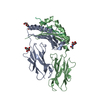 1iakS S: Starting model for refinement |
|---|---|
| Similar structure data |
-Links
 PDBj
PDBj


- Assembly
| Deposited unit | 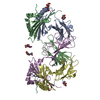
| ||||||||
|---|---|---|---|---|---|---|---|---|---|
| 1 |
| ||||||||
| Unit cell |
| ||||||||
| Components on special symmetry positions |
|
-Components
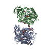
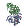
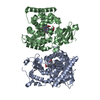
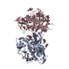
| #1: Protein | Mass: 20792.258 Da / Num. of mol.: 2 / Fragment: A-D ALPHA CHAIN Source method: isolated from a genetically manipulated source Source: (gene. exp.)   Spodoptera frugiperda (fall armyworm) / References: UniProt: P04228 Spodoptera frugiperda (fall armyworm) / References: UniProt: P04228#2: Protein | Mass: 22179.779 Da / Num. of mol.: 2 / Fragment: BETA CHAIN Source method: isolated from a genetically manipulated source Source: (gene. exp.) Spodoptera frugiperda (fall armyworm) / References: UniProt: Q31135#3: Protein/peptide | Mass: 1671.880 Da / Num. of mol.: 2 Fragment: RESIDUES 11-24, CORRESPOND TO BINDING SITES P-3 TO P11 Source method: isolated from a genetically manipulated source Source: (gene. exp.) Spodoptera frugiperda (fall armyworm) / References: UniProt: P00698#4: Sugar | ChemComp-NAG /   Type: D-saccharide, beta linking / Mass: 221.208 Da / Num. of mol.: 6 Type: D-saccharide, beta linking / Mass: 221.208 Da / Num. of mol.: 6Source method: isolated from a genetically manipulated source Formula: C8H15NO6 #5: Water | ChemComp-HOH / |  Mass: 18.015 Da / Num. of mol.: 72 / Source method: isolated from a natural source / Formula: H2O Mass: 18.015 Da / Num. of mol.: 72 / Source method: isolated from a natural source / Formula: H2OCompound details | THIS ENTRY CONTAINS COORDINATES FOR THE EXTRACELLULAR DOMAINS OF THE MURINE MHC CLASS II MOLECULE I- ...THIS ENTRY CONTAINS COORDINATE | Has protein modification | Y | |
|---|
-Experimental details
-Experiment
| Experiment | Method: X-RAY DIFFRACTION / Number of used crystals: 1 |
|---|
- Sample preparation
| Crystal | Density Matthews: 2.96 Å3/Da / Density % sol: 58.46 % | |||||||||||||||||||||||||||||||||||
|---|---|---|---|---|---|---|---|---|---|---|---|---|---|---|---|---|---|---|---|---|---|---|---|---|---|---|---|---|---|---|---|---|---|---|---|---|
| Crystal grow | pH: 4.4 / Details: pH 4.4 | |||||||||||||||||||||||||||||||||||
| Crystal grow | *PLUS Temperature: 20 ℃ / pH: 7.4 / Method: vapor diffusion, hanging drop | |||||||||||||||||||||||||||||||||||
| Components of the solutions | *PLUS
|
-Data collection
| Diffraction | Mean temperature: 100 K |
|---|---|
| Diffraction source | Source: ROTATING ANODE / Type: RIGAKU / Wavelength: 1.54 |
| Detector | Type: RIGAKU RAXIS IV / Detector: IMAGE PLATE / Details: YALE MIRRORS |
| Radiation | Protocol: SINGLE WAVELENGTH / Monochromatic (M) / Laue (L): M / Scattering type: x-ray |
| Radiation wavelength | Wavelength: 1.54 Å / Relative weight: 1 |
| Reflection | Resolution: 3.1→20 Å / Num. obs: 18859 / % possible obs: 93.7 % / Observed criterion σ(I): 0 / Redundancy: 5.5 % / Biso Wilson estimate: 39.1 Å2 / Rmerge(I) obs: 0.129 / Net I/σ(I): 6 |
| Reflection shell | Resolution: 3.1→3.29 Å / Rmerge(I) obs: 0.345 / Mean I/σ(I) obs: 2.3 / % possible all: 98.8 |
| Reflection | *PLUS Num. measured all: 102791 |
| Reflection shell | *PLUS % possible obs: 98.8 % / Num. unique obs: 3524 / Num. measured obs: 15897 |
- Processing
| Software |
| ||||||||||||||||||||||||||||||||||||||||||||||||||||||||||||||||||||||||||||||||
|---|---|---|---|---|---|---|---|---|---|---|---|---|---|---|---|---|---|---|---|---|---|---|---|---|---|---|---|---|---|---|---|---|---|---|---|---|---|---|---|---|---|---|---|---|---|---|---|---|---|---|---|---|---|---|---|---|---|---|---|---|---|---|---|---|---|---|---|---|---|---|---|---|---|---|---|---|---|---|---|---|---|
| Refinement | Method to determine structure: MOLECULAR REPLACEMENT Starting model: PDB ENTRY 1IAK Resolution: 3.1→19.82 Å / Rfactor Rfree error: 0.011 / Data cutoff high absF: 285984.27 / Data cutoff low absF: 0 / Isotropic thermal model: RESTRAINED / Cross valid method: THROUGHOUT / σ(F): 3
| ||||||||||||||||||||||||||||||||||||||||||||||||||||||||||||||||||||||||||||||||
| Displacement parameters | Biso mean: 36.4 Å2
| ||||||||||||||||||||||||||||||||||||||||||||||||||||||||||||||||||||||||||||||||
| Refine analyze |
| ||||||||||||||||||||||||||||||||||||||||||||||||||||||||||||||||||||||||||||||||
| Refinement step | Cycle: LAST / Resolution: 3.1→19.82 Å
| ||||||||||||||||||||||||||||||||||||||||||||||||||||||||||||||||||||||||||||||||
| Refine LS restraints |
| ||||||||||||||||||||||||||||||||||||||||||||||||||||||||||||||||||||||||||||||||
| LS refinement shell | Resolution: 3.1→3.29 Å / Rfactor Rfree error: 0.034 / Total num. of bins used: 6
| ||||||||||||||||||||||||||||||||||||||||||||||||||||||||||||||||||||||||||||||||
| Xplor file |
| ||||||||||||||||||||||||||||||||||||||||||||||||||||||||||||||||||||||||||||||||
| Software | *PLUS Name: CNS / Classification: refinement | ||||||||||||||||||||||||||||||||||||||||||||||||||||||||||||||||||||||||||||||||
| Refinement | *PLUS Highest resolution: 3.1 Å / σ(F): 3 / % reflection Rfree: 4.8 % | ||||||||||||||||||||||||||||||||||||||||||||||||||||||||||||||||||||||||||||||||
| Solvent computation | *PLUS | ||||||||||||||||||||||||||||||||||||||||||||||||||||||||||||||||||||||||||||||||
| Displacement parameters | *PLUS Biso mean: 36.4 Å2 | ||||||||||||||||||||||||||||||||||||||||||||||||||||||||||||||||||||||||||||||||
| Refine LS restraints | *PLUS
| ||||||||||||||||||||||||||||||||||||||||||||||||||||||||||||||||||||||||||||||||
| LS refinement shell | *PLUS Rfactor Rfree: 0.363 / % reflection Rfree: 5 % / Rfactor Rwork: 0.285 / Rfactor obs: 0.285 |
