| Software | | Name | Version | Classification |
|---|
| CBASS | | data collection| SHELXS | | phasing| PHENIX | (phenix.refine: 1.7.1_743)refinement| HKL-2000 | | data reduction| HKL-2000 | | data scaling | | | | | |
|
|---|
| Refinement | Method to determine structure:  SAD / Resolution: 3.187→35.518 Å / Occupancy max: 1 / Occupancy min: 1 / FOM work R set: 0.6955 / SU ML: 0.99 / σ(F): 1.35 / Phase error: 35.58 / Stereochemistry target values: ML SAD / Resolution: 3.187→35.518 Å / Occupancy max: 1 / Occupancy min: 1 / FOM work R set: 0.6955 / SU ML: 0.99 / σ(F): 1.35 / Phase error: 35.58 / Stereochemistry target values: ML
| Rfactor | Num. reflection | % reflection |
|---|
| Rfree | 0.3277 | 1146 | 5.13 % |
|---|
| Rwork | 0.3096 | 21209 | - |
|---|
| obs | 0.3106 | 22355 | 84.24 % |
|---|
|
|---|
| Solvent computation | Shrinkage radii: 0.38 Å / VDW probe radii: 0.7 Å / Solvent model: FLAT BULK SOLVENT MODEL / Bsol: 59.768 Å2 / ksol: 0.284 e/Å3 |
|---|
| Displacement parameters | Biso max: 257.79 Å2 / Biso mean: 108.5685 Å2 / Biso min: 20 Å2
| Baniso -1 | Baniso -2 | Baniso -3 |
|---|
| 1- | -19.8108 Å2 | -0 Å2 | -0 Å2 |
|---|
| 2- | - | -3.9388 Å2 | 0 Å2 |
|---|
| 3- | - | - | -4.8407 Å2 |
|---|
|
|---|
| Refinement step | Cycle: LAST / Resolution: 3.187→35.518 Å
| Protein | Nucleic acid | Ligand | Solvent | Total |
|---|
| Num. atoms | 7342 | 0 | 0 | 0 | 7342 |
|---|
|
|---|
| Refine LS restraints | | Refine-ID | Type | Dev ideal | Number |
|---|
| X-RAY DIFFRACTION | f_bond_d| 0.015 | 7527 | | X-RAY DIFFRACTION | f_angle_d| 1.64 | 10276 | | X-RAY DIFFRACTION | f_chiral_restr| 0.102 | 1257 | | X-RAY DIFFRACTION | f_plane_restr| 0.018 | 1245 | | X-RAY DIFFRACTION | f_dihedral_angle_d| 23.627 | 2563 | | | | | |
|
|---|
| LS refinement shell | Refine-ID: X-RAY DIFFRACTION / Total num. of bins used: 8 | Resolution (Å) | Rfactor Rfree | Num. reflection Rfree | Rfactor Rwork | Num. reflection Rwork | Num. reflection all | % reflection obs (%) |
|---|
| 3.1871-3.332 | 0.4199 | 50 | 0.4075 | 1036 | 1086 | 34 | | 3.332-3.5076 | 0.3921 | 107 | 0.3755 | 1858 | 1965 | 60 | | 3.5076-3.7271 | 0.395 | 129 | 0.3812 | 2549 | 2678 | 82 | | 3.7271-4.0145 | 0.3709 | 160 | 0.3644 | 3096 | 3256 | 99 | | 4.0145-4.4178 | 0.3262 | 158 | 0.3124 | 3141 | 3299 | 100 | | 4.4178-5.0555 | 0.3048 | 164 | 0.2661 | 3158 | 3322 | 100 | | 5.0555-6.3635 | 0.4031 | 190 | 0.3377 | 3145 | 3335 | 100 | | 6.3635-35.5199 | 0.2576 | 188 | 0.2646 | 3226 | 3414 | 97 |
|
|---|
| Refinement TLS params. | Method: refined / Origin x: -29.5593 Å / Origin y: -4.2023 Å / Origin z: 22.0175 Å
| 11 | 12 | 13 | 21 | 22 | 23 | 31 | 32 | 33 |
|---|
| T | 0.3221 Å2 | 0.1093 Å2 | -0.0556 Å2 | - | -0.0452 Å2 | 0.0661 Å2 | - | - | 0.2995 Å2 |
|---|
| L | 1.2984 °2 | 0.2871 °2 | -0.0397 °2 | - | 1.6562 °2 | 0.433 °2 | - | - | 7.6525 °2 |
|---|
| S | -0.0635 Å ° | 0.1598 Å ° | -0.0147 Å ° | -0.2188 Å ° | 0.0484 Å ° | 0.1916 Å ° | -0.8863 Å ° | -0.264 Å ° | -0.0126 Å ° |
|---|
|
|---|
| Refinement TLS group | | ID | Refine-ID | Refine TLS-ID | Selection details | Auth asym-ID | Auth seq-ID |
|---|
| 1 | X-RAY DIFFRACTION | 1 | allA| 10 - 509 | | 2 | X-RAY DIFFRACTION | 1 | allB| 12 - 504 | | | | |
|
|---|
 Movie
Movie Controller


 Open data
 Basic information
 Components
 Keywords
 Function and homology information
 Authors
 Citation
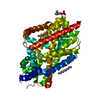
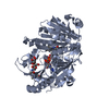
 Structure visualization
 Downloads & links
 Other downloads


 PDBj
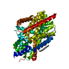
PDBj Assembly

 Sample preparation
 / Beamline: BL41XU / Wavelength: 1 Å
 Processing