 Movie
Movie Controller

[English] 日本語
 Yorodumi
- PDB-3sgh: Crystal structure of a SusD-like protein (BT_3752) from Bacteroid... -
+ Open data

- Basic information
| Entry | Database: PDB / ID: 3sgh | ||||||
|---|---|---|---|---|---|---|---|
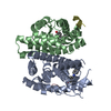
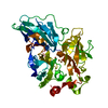
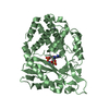
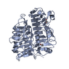
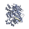
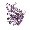
| Title | Crystal structure of a SusD-like protein (BT_3752) from Bacteroides thetaiotaomicron VPI-5482 at 1.70 A resolution | ||||||
 Components Components | SusD homolog | ||||||
 Keywords Keywords | SUGAR BINDING PROTEIN / alpha-alpha superhelix / Structural Genomics / Joint Center for Structural Genomics / JCSG / Protein Structure Initiative / PSI-BIOLOGY | ||||||
| Function / homology | SusD-like / Susd and RagB outer membrane lipoprotein / Serine Threonine Protein Phosphatase 5, Tetratricopeptide repeat - #390 / Serine Threonine Protein Phosphatase 5, Tetratricopeptide repeat / Alpha Horseshoe / Prokaryotic membrane lipoprotein lipid attachment site profile. / Tetratricopeptide-like helical domain superfamily / Mainly Alpha / SusD homolog Function and homology information Function and homology information | ||||||
| Biological species |  Bacteroides thetaiotaomicron (bacteria) Bacteroides thetaiotaomicron (bacteria) | ||||||
| Method |  X-RAY DIFFRACTION / SYNCHROTRON / MAD / Resolution: 1.7 Å X-RAY DIFFRACTION / SYNCHROTRON / MAD / Resolution: 1.7 Å | ||||||
 Authors Authors | Joint Center for Structural Genomics (JCSG) | ||||||
 Citation Citation | Journal: To be published Title: Crystal structure of a Hypothetical SusD-like protein (BT_3752) from Bacteroides thetaiotaomicron VPI-5482 at 1.70 A resolution Authors: Joint Center for Structural Genomics (JCSG) | ||||||
| History |
|
- Structure visualization
| Structure viewer | Molecule: MolmilJmol/JSmol |
|---|
- Downloads & links
-Download
| PDBx/mmCIF format | 3sgh.cif.gz | 409.6 KB | Display | PDBx/mmCIF format |
|---|---|---|---|---|
| PDB format | pdb3sgh.ent.gz | 334.4 KB | Display | PDB format |
| PDBx/mmJSON format | 3sgh.json.gz | Tree view | PDBx/mmJSON format | |
| Others |  Other downloads Other downloads |
-Validation report
| Arichive directory | https://data.pdbj.org/pub/pdb/validation_reports/sg/3sghftp://data.pdbj.org/pub/pdb/validation_reports/sg/3sgh | HTTPS FTP |
|---|
-Related structure data
| Similar structure data | |
|---|---|
| Other databases |


-Links
 PDBj
PDBj


- Assembly
| Deposited unit | 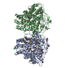
| ||||||||
|---|---|---|---|---|---|---|---|---|---|
| 1 | 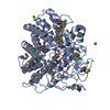
| ||||||||
| 2 | 
| ||||||||
| Unit cell |
| ||||||||
| Components on special symmetry positions |
|
-Components
| #1: Protein | Mass: 55942.598 Da / Num. of mol.: 2 / Fragment: sequence database residues 24-521 Source method: isolated from a genetically manipulated source Source: (gene. exp.) Bacteroides thetaiotaomicron (bacteria)Gene: BT_3752 / Plasmid: SpeedET / Production host: #2: Chemical | ChemComp-CL /   Mass: 35.453 Da / Num. of mol.: 20 / Source method: obtained synthetically / Formula: Cl Mass: 35.453 Da / Num. of mol.: 20 / Source method: obtained synthetically / Formula: Cl#3: Water | ChemComp-HOH / |  Mass: 18.015 Da / Num. of mol.: 1355 / Source method: isolated from a natural source / Formula: H2O Mass: 18.015 Da / Num. of mol.: 1355 / Source method: isolated from a natural source / Formula: H2OHas protein modification | Y | Sequence details | THIS CONSTRUCT (RESIDUES 24-521) WAS EXPRESSED WITH A PURIFICATION TAG MGSDKIHHHHHHENLYFQG. THE TAG ...THIS CONSTRUCT (RESIDUES 24-521) WAS EXPRESSED WITH A PURIFICATI | |
|---|
-Experimental details
-Experiment
| Experiment | Method: X-RAY DIFFRACTION / Number of used crystals: 1 |
|---|
- Sample preparation
| Crystal | Density Matthews: 2.17 Å3/Da / Density % sol: 43.37 % |
|---|---|
| Crystal grow | Temperature: 277 K / Method: vapor diffusion, sitting drop / pH: 6.5 Details: 20.00% polyethylene glycol 6000, 0.20M magnesium chloride, 0.1M sodium cacodylate pH 6.5, NANODROP, VAPOR DIFFUSION, SITTING DROP, temperature 277K |
-Data collection
| Diffraction | Mean temperature: 100 K | |||||||||||||||||||||||||||||||||||||||||||||||||||||||||||||||||||||||||||||
|---|---|---|---|---|---|---|---|---|---|---|---|---|---|---|---|---|---|---|---|---|---|---|---|---|---|---|---|---|---|---|---|---|---|---|---|---|---|---|---|---|---|---|---|---|---|---|---|---|---|---|---|---|---|---|---|---|---|---|---|---|---|---|---|---|---|---|---|---|---|---|---|---|---|---|---|---|---|---|
| Diffraction source | Source: SYNCHROTRON / Site: SSRL  / Beamline: BL11-1 / Wavelength: 0.91837,0.97944,0.97899 / Beamline: BL11-1 / Wavelength: 0.91837,0.97944,0.97899 | |||||||||||||||||||||||||||||||||||||||||||||||||||||||||||||||||||||||||||||
| Detector | Type: MARMOSAIC 325 mm CCD / Detector: CCD / Date: May 26, 2011 Details: Flat mirror (vertical focusing); single crystal Si(111) bent monochromator (ho rizontal focusing) | |||||||||||||||||||||||||||||||||||||||||||||||||||||||||||||||||||||||||||||
| Radiation | Monochromator: single crystal Si(111) bent / Protocol: MAD / Monochromatic (M) / Laue (L): M / Scattering type: x-ray | |||||||||||||||||||||||||||||||||||||||||||||||||||||||||||||||||||||||||||||
| Radiation wavelength |
| |||||||||||||||||||||||||||||||||||||||||||||||||||||||||||||||||||||||||||||
| Reflection | Resolution: 1.7→29.633 Å / Num. obs: 107089 / % possible obs: 97.1 % / Observed criterion σ(I): -3 / Biso Wilson estimate: 16.942 Å2 / Rmerge(I) obs: 0.089 / Net I/σ(I): 8.12 | |||||||||||||||||||||||||||||||||||||||||||||||||||||||||||||||||||||||||||||
| Reflection shell |
|
-Phasing
| Phasing | Method: MAD |
|---|
- Processing
| Software |
| |||||||||||||||||||||||||||||||||||||||||||||||||||||||||||||||||||||||||||||||||||||
|---|---|---|---|---|---|---|---|---|---|---|---|---|---|---|---|---|---|---|---|---|---|---|---|---|---|---|---|---|---|---|---|---|---|---|---|---|---|---|---|---|---|---|---|---|---|---|---|---|---|---|---|---|---|---|---|---|---|---|---|---|---|---|---|---|---|---|---|---|---|---|---|---|---|---|---|---|---|---|---|---|---|---|---|---|---|---|
| Refinement | Method to determine structure: MAD / Resolution: 1.7→29.633 Å / Cor.coef. Fo:Fc: 0.974 / Cor.coef. Fo:Fc free: 0.959 / Occupancy max: 1 / Occupancy min: 0.23 / SU B: 3.457 / SU ML: 0.059 / Cross valid method: THROUGHOUT / σ(F): 0 / ESU R: 0.09 / ESU R Free: 0.09 Stereochemistry target values: MAXIMUM LIKELIHOOD WITH PHASES Details: 1. HYDROGENS HAVE BEEN ADDED IN THE RIDING POSITIONS. 2. SOLVENT MOLECULES WERE NOT INCLUDED IN THE TLS GROUP DEFINITIONS. 3. ATOM RECORD CONTAINS SUM OF TLS AND RESIDUAL B FACTORS. 4. ...Details: 1. HYDROGENS HAVE BEEN ADDED IN THE RIDING POSITIONS. 2. SOLVENT MOLECULES WERE NOT INCLUDED IN THE TLS GROUP DEFINITIONS. 3. ATOM RECORD CONTAINS SUM OF TLS AND RESIDUAL B FACTORS. 4. ANISOU RECORD CONTAINS SUM OF TLS AND RESIDUAL U FACTORS. 5. A MET-INHIBITION PROTOCOL WAS USED FOR SELENOMETHIONINE INCORPORATION DURING PROTEIN EXPRESSION. THE OCCUPANCY OF THE SE ATOMS IN THE MSE RESIDUES WAS REDUCED TO 0.75 FOR THE REDUCED SCATTERING POWER DUE TO PARTIAL S-MET INCORPORATION. 6. CHLORIDE (CL) IONS MODELED ARE FROM CRYSTALLIZATION CONDITION.
| |||||||||||||||||||||||||||||||||||||||||||||||||||||||||||||||||||||||||||||||||||||
| Solvent computation | Ion probe radii: 0.8 Å / Shrinkage radii: 0.8 Å / VDW probe radii: 1.4 Å / Solvent model: BABINET MODEL WITH MASK | |||||||||||||||||||||||||||||||||||||||||||||||||||||||||||||||||||||||||||||||||||||
| Displacement parameters | Biso max: 57.98 Å2 / Biso mean: 20.9933 Å2 / Biso min: 10.49 Å2
| |||||||||||||||||||||||||||||||||||||||||||||||||||||||||||||||||||||||||||||||||||||
| Refinement step | Cycle: LAST / Resolution: 1.7→29.633 Å
| |||||||||||||||||||||||||||||||||||||||||||||||||||||||||||||||||||||||||||||||||||||
| Refine LS restraints |
| |||||||||||||||||||||||||||||||||||||||||||||||||||||||||||||||||||||||||||||||||||||
| LS refinement shell | Resolution: 1.7→1.744 Å / Total num. of bins used: 20
| |||||||||||||||||||||||||||||||||||||||||||||||||||||||||||||||||||||||||||||||||||||
| Refinement TLS params. | Method: refined / Refine-ID: X-RAY DIFFRACTION
| |||||||||||||||||||||||||||||||||||||||||||||||||||||||||||||||||||||||||||||||||||||
| Refinement TLS group |
|
