 Movie
Movie Controller

[English] 日本語
 Yorodumi
- PDB-4tyv: Ensemble refinement of the E502A variant of sacteLam55A from Stre... -
+ Open data

- Basic information
| Entry | Database: PDB / ID: 4tyv | |||||||||
|---|---|---|---|---|---|---|---|---|---|---|
| Title | Ensemble refinement of the E502A variant of sacteLam55A from Streptomyces sp. SirexAA-E in complex with glucose | |||||||||
 Components Components | Putative secreted protein | |||||||||
 Keywords Keywords | HYDROLASE / exo-beta-1 / 3-glucanase / beta-1 / GH55 / glucose / secreted / biomass degradation | |||||||||
| Function / homology | : / glucan 1,3-beta-glucosidase / glucan exo-1,3-beta-glucosidase activity / glucan catabolic process / Pectin lyase fold / extracellular region / beta-D-glucopyranose / Exo-beta-1,3-glucanase Function and homology information Function and homology information | |||||||||
| Biological species |  Streptomyces sp. SirexAA-E (bacteria) Streptomyces sp. SirexAA-E (bacteria) | |||||||||
| Method |  X-RAY DIFFRACTION / SYNCHROTRON / MOLECULAR REPLACEMENT / molecular replacement / Resolution: 1.75 Å X-RAY DIFFRACTION / SYNCHROTRON / MOLECULAR REPLACEMENT / molecular replacement / Resolution: 1.75 Å | |||||||||
 Authors Authors | Bianchetti, C.M. / Takasuka, T.E. / Yik, E.J. / Bergeman, L.F. / Fox, B.G. | |||||||||
 Citation Citation | Journal: J.Biol.Chem. / Year: 2015 Title: Active site and laminarin binding in glycoside hydrolase family 55. Authors: Bianchetti, C.M. / Takasuka, T.E. / Deutsch, S. / Udell, H.S. / Yik, E.J. / Bergeman, L.F. / Fox, B.G. | |||||||||
| History |
|
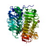
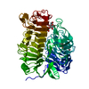
- Structure visualization
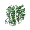
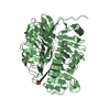
| Structure viewer | Molecule: MolmilJmol/JSmol |
|---|
- Downloads & links
-Download
| PDBx/mmCIF format | 4tyv.cif.gz | 17 MB | Display | PDBx/mmCIF format |
|---|---|---|---|---|
| PDB format | pdb4tyv.ent.gz | 14.5 MB | Display | PDB format |
| PDBx/mmJSON format | 4tyv.json.gz | Tree view | PDBx/mmJSON format | |
| Others |  Other downloads Other downloads |
-Validation report
| Arichive directory | https://data.pdbj.org/pub/pdb/validation_reports/ty/4tyvftp://data.pdbj.org/pub/pdb/validation_reports/ty/4tyv | HTTPS FTP |
|---|
-Related structure data
| Related structure data |  4pewSC 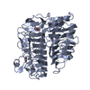 4pexC  4peyC  4pezC  4pf0C  4tz1C 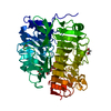 4tz3C  4tz5C C: citing same article ( S: Starting model for refinement |
|---|---|
| Similar structure data |


-Links
 PDBj
PDBj


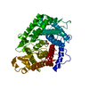
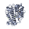
- Assembly
| Deposited unit | 
| ||||||||
|---|---|---|---|---|---|---|---|---|---|
| 1 | 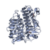
| ||||||||
| 2 | 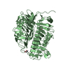
| ||||||||
| Unit cell |
| ||||||||
| Number of models | 25 |
-Components
| #1: Protein | Mass: 59083.562 Da / Num. of mol.: 2 Source method: isolated from a genetically manipulated source Source: (gene. exp.) Streptomyces sp. SirexAA-E (bacteria) / Gene: SACTE_4363 / Plasmid: PVP67K / Production host: #2: Chemical | ChemComp-EDO /   Mass: 62.068 Da / Num. of mol.: 5 / Source method: obtained synthetically / Formula: C2H6O2 Mass: 62.068 Da / Num. of mol.: 5 / Source method: obtained synthetically / Formula: C2H6O2#3: Sugar | ChemComp-BGC / |   Type: D-saccharide, beta linking / Mass: 180.156 Da / Num. of mol.: 1 Type: D-saccharide, beta linking / Mass: 180.156 Da / Num. of mol.: 1Source method: isolated from a genetically manipulated source Formula: C6H12O6 #4: Water | ChemComp-HOH / |  Mass: 18.015 Da / Num. of mol.: 503 / Source method: isolated from a natural source / Formula: H2O Mass: 18.015 Da / Num. of mol.: 503 / Source method: isolated from a natural source / Formula: H2O |
|---|
-Experimental details
-Experiment
| Experiment | Method: X-RAY DIFFRACTION / Number of used crystals: 1 |
|---|
- Sample preparation
| Crystal | Density Matthews: 2.27 Å3/Da / Density % sol: 45.85 % |
|---|---|
| Crystal grow | Temperature: 298 K / Method: vapor diffusion, hanging drop / pH: 6.5 Details: Protein Solution (20 mg/ml protein, 0.05 M NaCl, and 0.010 M MOPS pH 7) mixed in a 1:1 ratio with the Well Solution (20% PEG 3350, 125mM NaCH02, and 100mM BTP pH 6.5). Cryoprotected with 20% ...Details: Protein Solution (20 mg/ml protein, 0.05 M NaCl, and 0.010 M MOPS pH 7) mixed in a 1:1 ratio with the Well Solution (20% PEG 3350, 125mM NaCH02, and 100mM BTP pH 6.5). Cryoprotected with 20% PEG 3350, 125mM NaCH02, 75mM Glucose, 100mM BTP pH 6.5 and 15% ethylene glycol |
-Data collection
| Diffraction | Mean temperature: 100 K |
|---|---|
| Diffraction source | Source: SYNCHROTRON / Site: APS  / Beamline: 21-ID-G / Wavelength: 0.97857 Å / Beamline: 21-ID-G / Wavelength: 0.97857 Å |
| Detector | Type: MARMOSAIC 300 mm CCD / Detector: CCD / Date: Aug 8, 2013 / Details: mirrors and beryllium lenses |
| Radiation | Monochromator: C(111) / Protocol: SINGLE WAVELENGTH / Monochromatic (M) / Laue (L): M / Scattering type: x-ray |
| Radiation wavelength | Wavelength: 0.97857 Å / Relative weight: 1 |
| Reflection | Resolution: 1.66→50 Å / Num. obs: 123726 / % possible obs: 99.9 % / Redundancy: 3.7 % / Rmerge(I) obs: 0.082 / Net I/σ(I): 7.2 |
| Reflection shell | Resolution: 1.66→1.69 Å / Redundancy: 3.5 % / Rmerge(I) obs: 0.546 / % possible all: 100 |
-Phasing
| Phasing | Method: molecular replacement |
|---|
- Processing
| Software |
| |||||||||||||||||||||||||||||||||||||||||||||||||||||||||||||||||||||||||||||||||||||||||||||||||||||||||||||||||||||||||||||||||||||||||||||||||||||||||||||||||||||||||||||||||||||||||||||||||||||||||||||||||||||||||
|---|---|---|---|---|---|---|---|---|---|---|---|---|---|---|---|---|---|---|---|---|---|---|---|---|---|---|---|---|---|---|---|---|---|---|---|---|---|---|---|---|---|---|---|---|---|---|---|---|---|---|---|---|---|---|---|---|---|---|---|---|---|---|---|---|---|---|---|---|---|---|---|---|---|---|---|---|---|---|---|---|---|---|---|---|---|---|---|---|---|---|---|---|---|---|---|---|---|---|---|---|---|---|---|---|---|---|---|---|---|---|---|---|---|---|---|---|---|---|---|---|---|---|---|---|---|---|---|---|---|---|---|---|---|---|---|---|---|---|---|---|---|---|---|---|---|---|---|---|---|---|---|---|---|---|---|---|---|---|---|---|---|---|---|---|---|---|---|---|---|---|---|---|---|---|---|---|---|---|---|---|---|---|---|---|---|---|---|---|---|---|---|---|---|---|---|---|---|---|---|---|---|---|---|---|---|---|---|---|---|---|---|---|---|---|---|---|---|---|
| Refinement | Method to determine structure: MOLECULAR REPLACEMENT Starting model: 4PEW Resolution: 1.75→24.27 Å / SU ML: 0.07 / Cross valid method: FREE R-VALUE / σ(F): 1.35 / Phase error: 14.57 / Stereochemistry target values: ML
| |||||||||||||||||||||||||||||||||||||||||||||||||||||||||||||||||||||||||||||||||||||||||||||||||||||||||||||||||||||||||||||||||||||||||||||||||||||||||||||||||||||||||||||||||||||||||||||||||||||||||||||||||||||||||
| Solvent computation | Shrinkage radii: 0.8 Å / VDW probe radii: 1 Å / Solvent model: FLAT BULK SOLVENT MODEL | |||||||||||||||||||||||||||||||||||||||||||||||||||||||||||||||||||||||||||||||||||||||||||||||||||||||||||||||||||||||||||||||||||||||||||||||||||||||||||||||||||||||||||||||||||||||||||||||||||||||||||||||||||||||||
| Refinement step | Cycle: LAST / Resolution: 1.75→24.27 Å
| |||||||||||||||||||||||||||||||||||||||||||||||||||||||||||||||||||||||||||||||||||||||||||||||||||||||||||||||||||||||||||||||||||||||||||||||||||||||||||||||||||||||||||||||||||||||||||||||||||||||||||||||||||||||||
| LS refinement shell |
|
