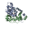
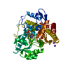
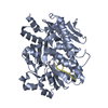
[English] 日本語
Yorodumi
- PDB-5bud: Crystal structure of Candida albicans Rai1 in complex with pU5-Mn2+ -

+
Open data


ID or keywords:

Loading...

-
Basic information

Entry
Database: PDB / ID: 5bud
TitleCrystal structure of Candida albicans Rai1 in complex with pU5-Mn2+
Components
  • Decapping nuclease RAI1
  • RNA (5'-R(P*UP*UP*UP*U)-3')
Keywordshydrolase/RNA / Rai1 / RNA / Decapping / mRNA 5'-processing / hydrolase-RNA complex
Function / homology
Function and homology information


RNA polymerase II termination complex / positive regulation of termination of RNA polymerase II transcription / RNA NAD+-cap (NAD+-forming) hydrolase activity / Las1 complex / termination of RNA polymerase II transcription, poly(A)-coupled / phosphodiesterase decapping endonuclease activity / deadenylation-independent decapping of nuclear-transcribed mRNA / mRNA 5'-diphosphatase activity / nuclear polyadenylation-dependent rRNA catabolic process / NAD-cap decapping ...RNA polymerase II termination complex / positive regulation of termination of RNA polymerase II transcription / RNA NAD+-cap (NAD+-forming) hydrolase activity / Las1 complex / termination of RNA polymerase II transcription, poly(A)-coupled / phosphodiesterase decapping endonuclease activity / deadenylation-independent decapping of nuclear-transcribed mRNA / mRNA 5'-diphosphatase activity / nuclear polyadenylation-dependent rRNA catabolic process / NAD-cap decapping / : / nuclear-transcribed mRNA catabolic process / cleavage in ITS2 between 5.8S rRNA and LSU-rRNA of tricistronic rRNA transcript (SSU-rRNA, 5.8S rRNA, LSU-rRNA) / Hydrolases; Acting on acid anhydrides; In phosphorus-containing anhydrides / enzyme regulator activity / mRNA processing / nucleotide binding / RNA binding / metal ion binding / nucleus / cytosol
Similarity search - Function
RAI1-like / RAI1 like PD-(D/E)XK nuclease / RAI1-like family
Similarity search - Domain/homology
: / RNA / Decapping nuclease RAI1
Similarity search - Component
Biological speciesCandida albicans (yeast)
MethodX-RAY DIFFRACTION / SYNCHROTRON / FOURIER SYNTHESIS / Resolution: 1.99 Å
AuthorsWang, V.Y. / Tong, L.
Funding support United States, 1items
OrganizationGrant numberCountry
National Institutes of Health/National Institute of General Medical Sciences (NIH/NIGMS)GM090059 United States
CitationJournal: Nucleic Acids Res. / Year: 2015
Title: Structural and biochemical studies of the distinct activity profiles of Rai1 enzymes.
Authors: Wang, V.Y. / Jiao, X. / Kiledjian, M. / Tong, L.
History
DepositionJun 3, 2015Deposition site: RCSB / Processing site: RCSB
Revision 1.0Jul 8, 2015Provider: repository / Type: Initial release
Revision 1.1Aug 5, 2015Group: Database references
Revision 1.2Sep 20, 2017Group: Author supporting evidence / Database references / Derived calculations
Category: citation / pdbx_audit_support / pdbx_struct_oper_list
Item: _citation.journal_id_CSD / _pdbx_audit_support.funding_organization / _pdbx_struct_oper_list.symmetry_operation
Revision 1.3Dec 25, 2019Group: Author supporting evidence / Category: pdbx_audit_support / Item: _pdbx_audit_support.funding_organization
Revision 1.4Mar 6, 2024Group: Data collection / Database references / Derived calculations
Category: chem_comp_atom / chem_comp_bond ...chem_comp_atom / chem_comp_bond / database_2 / pdbx_struct_conn_angle / struct_conn
Item: _database_2.pdbx_DOI / _database_2.pdbx_database_accession ..._database_2.pdbx_DOI / _database_2.pdbx_database_accession / _pdbx_struct_conn_angle.ptnr1_auth_asym_id / _pdbx_struct_conn_angle.ptnr1_auth_comp_id / _pdbx_struct_conn_angle.ptnr1_auth_seq_id / _pdbx_struct_conn_angle.ptnr1_label_asym_id / _pdbx_struct_conn_angle.ptnr1_label_atom_id / _pdbx_struct_conn_angle.ptnr1_label_comp_id / _pdbx_struct_conn_angle.ptnr1_label_seq_id / _pdbx_struct_conn_angle.ptnr2_auth_seq_id / _pdbx_struct_conn_angle.ptnr2_label_asym_id / _pdbx_struct_conn_angle.ptnr3_auth_asym_id / _pdbx_struct_conn_angle.ptnr3_auth_comp_id / _pdbx_struct_conn_angle.ptnr3_auth_seq_id / _pdbx_struct_conn_angle.ptnr3_label_asym_id / _pdbx_struct_conn_angle.ptnr3_label_atom_id / _pdbx_struct_conn_angle.ptnr3_label_comp_id / _pdbx_struct_conn_angle.ptnr3_label_seq_id / _pdbx_struct_conn_angle.value / _struct_conn.pdbx_dist_value / _struct_conn.ptnr1_auth_asym_id / _struct_conn.ptnr1_auth_comp_id / _struct_conn.ptnr1_auth_seq_id / _struct_conn.ptnr1_label_asym_id / _struct_conn.ptnr1_label_atom_id / _struct_conn.ptnr1_label_comp_id / _struct_conn.ptnr1_label_seq_id / _struct_conn.ptnr2_auth_asym_id / _struct_conn.ptnr2_auth_comp_id / _struct_conn.ptnr2_auth_seq_id / _struct_conn.ptnr2_label_asym_id / _struct_conn.ptnr2_label_atom_id / _struct_conn.ptnr2_label_comp_id / _struct_conn.ptnr2_label_seq_id

-
Structure visualization

Structure viewerMolecule:
MolmilJmol/JSmol

Downloads & links

-
Assembly

Deposited unit
A: Decapping nuclease RAI1
B: Decapping nuclease RAI1
D: RNA (5'-R(P*UP*UP*UP*U)-3')
E: RNA (5'-R(P*UP*UP*UP*U)-3')
hetero molecules


Theoretical massNumber of molelcules
Total (without water)95,07010
Polymers94,7404
Non-polymers3306
Water18,8081044
1
A: Decapping nuclease RAI1
D: RNA (5'-R(P*UP*UP*UP*U)-3')
hetero molecules


Theoretical massNumber of molelcules
Total (without water)47,5355
Polymers47,3702
Non-polymers1653
Water362
TypeNameSymmetry operationNumber
identity operation1_555x,y,z1
Buried area1770 Å2
ΔGint-35 kcal/mol
Surface area19440 Å2
MethodPISA
2
B: Decapping nuclease RAI1
E: RNA (5'-R(P*UP*UP*UP*U)-3')
hetero molecules


Theoretical massNumber of molelcules
Total (without water)47,5355
Polymers47,3702
Non-polymers1653
Water362
TypeNameSymmetry operationNumber
identity operation1_555x,y,z1
Buried area1880 Å2
ΔGint-36 kcal/mol
Surface area19280 Å2
MethodPISA
Unit cell
Length a, b, c (Å)97.854, 114.638, 84.458
Angle α, β, γ (deg.)90.00, 90.00, 90.00
Int Tables number18
Space group name H-MP21212

-
Components

#1: Protein Decapping nuclease RAI1


Mass: 45884.219 Da / Num. of mol.: 2
Source method: isolated from a genetically manipulated source
Source: (gene. exp.) Candida albicans (strain SC5314 / ATCC MYA-2876) (yeast)
Strain: SC5314 / ATCC MYA-2876 / Gene: RAI1, CaO19.13610, CaO19.6230 / Production host: Escherichia coli (E. coli)
References: UniProt: Q5AAT0, Hydrolases; Acting on acid anhydrides; In phosphorus-containing anhydrides
#2: RNA chain RNA (5'-R(P*UP*UP*UP*U)-3')


Mass: 1485.872 Da / Num. of mol.: 2 / Source method: obtained synthetically / Source: (synth.) Candida albicans (yeast)
#3: Chemical
ChemComp-MN / MANGANESE (II) ION


Mass: 54.938 Da / Num. of mol.: 6 / Source method: obtained synthetically / Formula: Mn
#4: Water ChemComp-HOH / water / Water


Mass: 18.015 Da / Num. of mol.: 1044 / Source method: isolated from a natural source / Formula: H2O

-
Experimental details

-
Experiment

ExperimentMethod: X-RAY DIFFRACTION

-
Sample preparation

CrystalDensity Matthews: 2.5 Å3/Da / Density % sol: 50.87 %
Crystal growTemperature: 293 K / Method: vapor diffusion, sitting drop
Details: CaRai1-pU5-Mn2+ complex crystals were obtained using 1:2 protein:RNA molar ratio and 10mM MnCl2 in 0.1M Bis-Tris (pH 5.5) and 18% (w/v) PEG3350

-
Data collection

DiffractionMean temperature: 100 K
Diffraction sourceSource: SYNCHROTRON / Site: NSLS / Beamline: X29A / Wavelength: 1 Å
DetectorType: ADSC QUANTUM 315r / Detector: CCD / Date: Feb 25, 2014
RadiationMonochromator: Si / Protocol: SINGLE WAVELENGTH / Monochromatic (M) / Laue (L): M / Scattering type: x-ray
Radiation wavelengthWavelength: 1 Å / Relative weight: 1
ReflectionResolution: 1.99→50 Å / Num. obs: 61906 / % possible obs: 99 % / Redundancy: 6.9 % / Rmerge(I) obs: 0.09 / Net I/σ(I): 19.6
Reflection shellResolution: 2→2.07 Å / Redundancy: 6.5 % / Rmerge(I) obs: 0.422 / Mean I/σ(I) obs: 4.2 / % possible all: 99

-
Processing

Software
NameVersionClassification
REFMAC5.7.0029refinement
HKL-2000data reduction
HKL-2000data scaling
PHASERphasing
RefinementMethod to determine structure: FOURIER SYNTHESIS / Resolution: 1.99→47.47 Å / Cor.coef. Fo:Fc: 0.957 / Cor.coef. Fo:Fc free: 0.932 / SU B: 3.877 / SU ML: 0.11 / Cross valid method: THROUGHOUT / ESU R: 0.187 / ESU R Free: 0.173 / Stereochemistry target values: MAXIMUM LIKELIHOOD / Details: HYDROGENS HAVE BEEN ADDED IN THE RIDING POSITIONS
RfactorNum. reflection% reflectionSelection details
Rfree0.24301 3301 5.1 %RANDOM
Rwork0.18651 ---
obs0.18947 61906 99.2 %-
Solvent computationIon probe radii: 0.8 Å / Shrinkage radii: 0.8 Å / VDW probe radii: 1.2 Å / Solvent model: MASK
Displacement parametersBiso mean: 33.431 Å2
Baniso -1Baniso -2Baniso -3
1-0.54 Å2-0 Å20 Å2
2---1.77 Å2-0 Å2
3---1.23 Å2
Refinement stepCycle: 1 / Resolution: 1.99→47.47 Å
ProteinNucleic acidLigandSolventTotal
Num. atoms6405 166 6 1044 7621
Refine LS restraints
Refine-IDTypeDev idealDev ideal targetNumber
X-RAY DIFFRACTIONr_bond_refined_d0.010.0196711
X-RAY DIFFRACTIONr_bond_other_d0.0010.026343
X-RAY DIFFRACTIONr_angle_refined_deg1.3961.9529100
X-RAY DIFFRACTIONr_angle_other_deg0.803314674
X-RAY DIFFRACTIONr_dihedral_angle_1_deg5.4465775
X-RAY DIFFRACTIONr_dihedral_angle_2_deg35.78524.984317
X-RAY DIFFRACTIONr_dihedral_angle_3_deg15.856151249
X-RAY DIFFRACTIONr_dihedral_angle_4_deg12.451536
X-RAY DIFFRACTIONr_chiral_restr0.0830.21007
X-RAY DIFFRACTIONr_gen_planes_refined0.0060.027341
X-RAY DIFFRACTIONr_gen_planes_other0.0010.021481
X-RAY DIFFRACTIONr_nbd_refined
X-RAY DIFFRACTIONr_nbd_other
X-RAY DIFFRACTIONr_nbtor_refined
X-RAY DIFFRACTIONr_nbtor_other
X-RAY DIFFRACTIONr_xyhbond_nbd_refined
X-RAY DIFFRACTIONr_xyhbond_nbd_other
X-RAY DIFFRACTIONr_metal_ion_refined
X-RAY DIFFRACTIONr_metal_ion_other
X-RAY DIFFRACTIONr_symmetry_vdw_refined
X-RAY DIFFRACTIONr_symmetry_vdw_other
X-RAY DIFFRACTIONr_symmetry_hbond_refined
X-RAY DIFFRACTIONr_symmetry_hbond_other
X-RAY DIFFRACTIONr_symmetry_metal_ion_refined
X-RAY DIFFRACTIONr_symmetry_metal_ion_other
X-RAY DIFFRACTIONr_mcbond_it
X-RAY DIFFRACTIONr_mcbond_other
X-RAY DIFFRACTIONr_mcangle_it
X-RAY DIFFRACTIONr_mcangle_other
X-RAY DIFFRACTIONr_scbond_it
X-RAY DIFFRACTIONr_scbond_other
X-RAY DIFFRACTIONr_scangle_it
X-RAY DIFFRACTIONr_scangle_other
X-RAY DIFFRACTIONr_long_range_B_refined
X-RAY DIFFRACTIONr_long_range_B_other
X-RAY DIFFRACTIONr_rigid_bond_restr
X-RAY DIFFRACTIONr_sphericity_free
X-RAY DIFFRACTIONr_sphericity_bonded
LS refinement shellResolution: 1.991→2.099 Å / Total num. of bins used: 10
RfactorNum. reflection% reflection
Rfree0.267 460 -
Rwork0.21 8631 -
obs--96.26 %

+
About Yorodumi

-
News

-
Feb 9, 2022. New format data for meta-information of EMDB entries

New format data for meta-information of EMDB entries

  • Version 3 of the EMDB header file is now the official format.
  • The previous official version 1.9 will be removed from the archive.

Related info.:EMDB header

External links:wwPDB to switch to version 3 of the EMDB data model

-
Aug 12, 2020. Covid-19 info

Covid-19 info

URL: https://pdbj.org/emnavi/covid19.php

New page: Covid-19 featured information page in EM Navigator.

Related info.:Covid-19 info / Mar 5, 2020. Novel coronavirus structure data

+
Mar 5, 2020. Novel coronavirus structure data

Novel coronavirus structure data

Related info.:Yorodumi Speices / Aug 12, 2020. Covid-19 info

External links:COVID-19 featured content - PDBj / Molecule of the Month (242):Coronavirus Proteases

+
Jan 31, 2019. EMDB accession codes are about to change! (news from PDBe EMDB page)

EMDB accession codes are about to change! (news from PDBe EMDB page)

  • The allocation of 4 digits for EMDB accession codes will soon come to an end. Whilst these codes will remain in use, new EMDB accession codes will include an additional digit and will expand incrementally as the available range of codes is exhausted. The current 4-digit format prefixed with “EMD-” (i.e. EMD-XXXX) will advance to a 5-digit format (i.e. EMD-XXXXX), and so on. It is currently estimated that the 4-digit codes will be depleted around Spring 2019, at which point the 5-digit format will come into force.
  • The EM Navigator/Yorodumi systems omit the EMD- prefix.

Related info.:Q: What is EMD? / ID/Accession-code notation in Yorodumi/EM Navigator

External links:EMDB Accession Codes are Changing Soon! / Contact to PDBj

+
Jul 12, 2017. Major update of PDB

Major update of PDB

  • wwPDB released updated PDB data conforming to the new PDBx/mmCIF dictionary.
  • This is a major update changing the version number from 4 to 5, and with Remediation, in which all the entries are updated.
  • In this update, many items about electron microscopy experimental information are reorganized (e.g. em_software).
  • Now, EM Navigator and Yorodumi are based on the updated data.

External links:wwPDB Remediation / Enriched Model Files Conforming to OneDep Data Standards Now Available in the PDB FTP Archive

-
Yorodumi

Thousand views of thousand structures

  • Yorodumi is a browser for structure data from EMDB, PDB, SASBDB, etc.
  • This page is also the successor to EM Navigator detail page, and also detail information page/front-end page for Omokage search.
  • The word "yorodu" (or yorozu) is an old Japanese word meaning "ten thousand". "mi" (miru) is to see.

Related info.:EMDB / PDB / SASBDB / Comparison of 3 databanks / Yorodumi Search / Aug 31, 2016. New EM Navigator & Yorodumi / Yorodumi Papers / Jmol/JSmol / Function and homology information / Changes in new EM Navigator and Yorodumi

Read more