Mass: 18.015 Da / Num. of mol.: 1232 / Source method: isolated from a natural source / Formula: H2O
Has protein modification
Y
Sequence details
THE CONSTRUCT (RESIDUES 28-480) WAS EXPRESSED WITH AN N-TERMINAL PURIFICATION TAG ...THE CONSTRUCT (RESIDUES 28-480) WAS EXPRESSED WITH AN N-TERMINAL PURIFICATION TAG MGSDKIHHHHHHENLYFQG. THE TAG WAS REMOVED WITH TEV PROTEASE LEAVING ONLY A GLYCINE (0) FOLLOWED BY THE TARGET SEQUENCE.
-
Experimental details
-
Experiment
Experiment
Method: X-RAY DIFFRACTION / Number of used crystals: 1
-
Sample preparation
Crystal
Density Matthews: 2.5 Å3/Da / Density % sol: 50.74 %
Resolution: 1.8→74.434 Å / Num. obs: 96603 / % possible obs: 99.9 % / Redundancy: 3.7 % / Biso Wilson estimate: 18.505 Å2 / Rsym value: 0.112 / Net I/σ(I): 6.4
Reflection shell
Resolution (Å)
Redundancy (%)
Rmerge(I) obs
Mean I/σ(I) obs
Num. measured all
Num. unique all
Rsym value
% possible all
1.8-1.9
3.7
0.59
1.9
51786
13961
0.59
99.9
1.9-2.01
3.7
0.412
2.6
48873
13163
0.412
100
2.01-2.15
3.7
0.272
3.8
46407
12468
0.272
100
2.15-2.32
3.7
0.2
4.8
43356
11635
0.2
100
2.32-2.55
3.7
0.149
6.2
39910
10712
0.149
100
2.55-2.85
3.7
0.113
7.7
36199
9724
0.113
100
2.85-3.29
3.7
0.085
9.9
32055
8631
0.085
100
3.29-4.02
3.7
0.066
13.4
27234
7344
0.066
99.8
4.02-5.69
3.7
0.058
14.5
21062
5731
0.058
99.5
5.69-74.43
3.5
0.063
13.4
11478
3234
0.063
97.8
-
Phasing
Phasing
Method: MAD
-
Processing
Software
Name
Version
Classification
NB
REFMAC
5.5.0109
refinement
PHENIX
refinement
SOLVE
phasing
MolProbity
3beta29
modelbuilding
SCALA
3.3.15
datascaling
PDB_EXTRACT
3.006
dataextraction
MOSFLM
datareduction
Refinement
Method to determine structure: MAD / Resolution: 1.8→74.434 Å / Cor.coef. Fo:Fc: 0.967 / Cor.coef. Fo:Fc free: 0.953 / Occupancy max: 1 / Occupancy min: 0.37 / SU B: 4.355 / SU ML: 0.071 / Cross valid method: THROUGHOUT / σ(F): 0 / ESU R Free: 0.106 Stereochemistry target values: MAXIMUM LIKELIHOOD WITH PHASES Details: 1. HYDROGENS HAVE BEEN ADDED IN THE RIDING POSITIONS. 2. A MET-INHIBITION PROTOCOL WAS USED FOR SELENOMETHIONINE INCORPORATION DURING PROTEIN EXPRESSION. THE OCCUPANCY OF THE SE ATOMS IN THE ...Details: 1. HYDROGENS HAVE BEEN ADDED IN THE RIDING POSITIONS. 2. A MET-INHIBITION PROTOCOL WAS USED FOR SELENOMETHIONINE INCORPORATION DURING PROTEIN EXPRESSION. THE OCCUPANCY OF THE SE ATOMS IN THE MSE RESIDUES WAS REDUCED TO 0.75 FOR THE REDUCED SCATTERING POWER DUE TO PARTIAL S-MET INCORPORATION. 3. ATOM RECORD CONTAINS SUM OF TLS AND RESIDUAL B FACTORS. ANISOU RECORD CONTAINS SUM OF TLS AND RESIDUAL U FACTORS. 4. WATERS WERE EXCLUDED FROM TLS GROUPS. 5. RAMACHANDRAN OUTLIERS (A61 AND B61) ARE SUPPORTED BY CLEAR DENSITY. 5. SODIUM ION (NA), PHOSPHATE ION (PO4), AND ETHYLENE GLYCOL (EDO) MODELED WERE PRESENT IN CRYSTLLIZATION/CRYO CONDITIONS.
Rfactor
Num. reflection
% reflection
Selection details
Rfree
0.19
4830
5 %
RANDOM
Rwork
0.151
-
-
-
obs
0.153
96490
99.74 %
-
Solvent computation
Ion probe radii: 0.8 Å / Shrinkage radii: 0.8 Å / VDW probe radii: 1.2 Å / Solvent model: MASK
In the structure databanks used in Yorodumi, some data are registered as the other names, "COVID-19 virus" and "2019-nCoV". Here are the details of the virus and the list of structure data.
Jan 31, 2019. EMDB accession codes are about to change! (news from PDBe EMDB page)
EMDB accession codes are about to change! (news from PDBe EMDB page)
The allocation of 4 digits for EMDB accession codes will soon come to an end. Whilst these codes will remain in use, new EMDB accession codes will include an additional digit and will expand incrementally as the available range of codes is exhausted. The current 4-digit format prefixed with “EMD-” (i.e. EMD-XXXX) will advance to a 5-digit format (i.e. EMD-XXXXX), and so on. It is currently estimated that the 4-digit codes will be depleted around Spring 2019, at which point the 5-digit format will come into force.
The EM Navigator/Yorodumi systems omit the EMD- prefix.
Related info.:Q: What is EMD? / ID/Accession-code notation in Yorodumi/EM Navigator
Yorodumi is a browser for structure data from EMDB, PDB, SASBDB, etc.
This page is also the successor to EM Navigator detail page, and also detail information page/front-end page for Omokage search.
The word "yorodu" (or yorozu) is an old Japanese word meaning "ten thousand". "mi" (miru) is to see.
Related info.:EMDB / PDB / SASBDB / Comparison of 3 databanks / Yorodumi Search / Aug 31, 2016. New EM Navigator & Yorodumi / Yorodumi Papers / Jmol/JSmol / Function and homology information / Changes in new EM Navigator and Yorodumi
 Movie
Movie Controller

 Yorodumi
 Open data
 Basic information
 Components
 Keywords
 Function and homology information
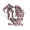
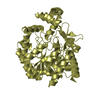
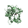
 Bacteroides vulgatus (bacteria)
 X-RAY DIFFRACTION /
  Authors
 Citation
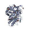
 Structure visualization
 Downloads & links
 Other downloads
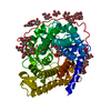


 PDBj
PDBj


 Assembly
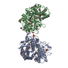


 Mass: 22.990 Da / Num. of mol.: 1 / Source method: obtained synthetically / Formula: Na
 Mass: 62.068 Da / Num. of mol.: 18 / Source method: obtained synthetically / Formula: C2H6O2
 Mass: 94.971 Da / Num. of mol.: 1 / Source method: obtained synthetically / Formula: PO4
 Mass: 18.015 Da / Num. of mol.: 1232 / Source method: isolated from a natural source / Formula: H2O
 Sample preparation
 / Beamline: BL9-2 / Wavelength: 0.91837,0.97954,0.97934
 Processing