 Movie
Movie Controller

[English] 日本語
 Yorodumi
- PDB-2hxu: Crystal structure of K220A mutant of L-Fuconate Dehydratase from ... -
+ Open data

- Basic information
| Entry | Database: PDB / ID: 2hxu | |||||||||
|---|---|---|---|---|---|---|---|---|---|---|
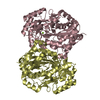
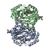
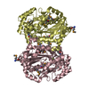
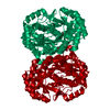
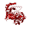
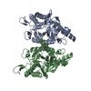
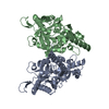
| Title | Crystal structure of K220A mutant of L-Fuconate Dehydratase from Xanthomonas campestris liganded with Mg++ and L-fuconate | |||||||||
 Components Components | L-fuconate dehydratase | |||||||||
 Keywords Keywords | UNKNOWN FUNCTION / L-fuconate dehydratase / enolase superfamily / L-fuconate | |||||||||
| Function / homology |  Function and homology information Function and homology informationL-fuconate dehydratase / L-fuconate dehydratase activity / hydro-lyase activity / carbohydrate catabolic process / magnesium ion binding Similarity search - Function | |||||||||
| Biological species |  Xanthomonas campestris pv. campestris (bacteria) Xanthomonas campestris pv. campestris (bacteria) | |||||||||
| Method |  X-RAY DIFFRACTION / SYNCHROTRON / MOLECULAR REPLACEMENT / Resolution: 1.8 Å X-RAY DIFFRACTION / SYNCHROTRON / MOLECULAR REPLACEMENT / Resolution: 1.8 Å | |||||||||
 Authors Authors | Fedorov, A.A. / Fedorov, E.V. / Yew, W.S. / Rakus, J.F. / Gerlt, J.A. / Almo, S.C. | |||||||||
 Citation Citation | Journal: Biochemistry / Year: 2006 Title: Evolution of Enzymatic Activities in the Enolase Superfamily: l-Fuconate Dehydratase from Xanthomonas campestris. Authors: Yew, W.S. / Fedorov, A.A. / Fedorov, E.V. / Rakus, J.F. / Pierce, R.W. / Almo, S.C. / Gerlt, J.A. | |||||||||
| History |
|
- Structure visualization
| Structure viewer | Molecule: MolmilJmol/JSmol |
|---|
- Downloads & links
-Download
| PDBx/mmCIF format | 2hxu.cif.gz | 105.5 KB | Display | PDBx/mmCIF format |
|---|---|---|---|---|
| PDB format | pdb2hxu.ent.gz | 78.9 KB | Display | PDB format |
| PDBx/mmJSON format | 2hxu.json.gz | Tree view | PDBx/mmJSON format | |
| Others |  Other downloads Other downloads |
-Validation report
| Arichive directory | https://data.pdbj.org/pub/pdb/validation_reports/hx/2hxuftp://data.pdbj.org/pub/pdb/validation_reports/hx/2hxu | HTTPS FTP |
|---|
-Related structure data
| Related structure data |  2hxtC 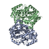 2hneS S: Starting model for refinement C: citing same article ( |
|---|---|
| Similar structure data |
-Links
 PDBj
PDBj
- Assembly
| Deposited unit | 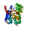
| ||||||||
|---|---|---|---|---|---|---|---|---|---|
| 1 | 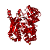
| ||||||||
| Unit cell |
| ||||||||
| Details | The biological assembly is a dimer, generated from the monomer in the asymmetric unit by crystallographic two fold axis |
-Components
| #1: Protein | Mass: 48120.516 Da / Num. of mol.: 1 Source method: isolated from a genetically manipulated source Source: (gene. exp.) Xanthomonas campestris pv. campestris (bacteria)Species: Xanthomonas campestris / Strain: ATCC 33913 / Production host: | ||||
|---|---|---|---|---|---|
| #2: Chemical | ChemComp-MG /   Mass: 24.305 Da / Num. of mol.: 1 / Source method: obtained synthetically / Formula: Mg Mass: 24.305 Da / Num. of mol.: 1 / Source method: obtained synthetically / Formula: Mg | ||||
| #3: Chemical |   Mass: 96.063 Da / Num. of mol.: 3 / Source method: obtained synthetically / Formula: SO4 Mass: 96.063 Da / Num. of mol.: 3 / Source method: obtained synthetically / Formula: SO4#4: Sugar | ChemComp-LFC / |   Type: L-saccharide / Mass: 180.156 Da / Num. of mol.: 1 Type: L-saccharide / Mass: 180.156 Da / Num. of mol.: 1Source method: isolated from a genetically manipulated source Formula: C6H12O6 #5: Water | ChemComp-HOH / |  Mass: 18.015 Da / Num. of mol.: 355 / Source method: isolated from a natural source / Formula: H2O Mass: 18.015 Da / Num. of mol.: 355 / Source method: isolated from a natural source / Formula: H2O |
-Experimental details
-Experiment
| Experiment | Method: X-RAY DIFFRACTION / Number of used crystals: 1 |
|---|
- Sample preparation
| Crystal | Density Matthews: 3.87 Å3/Da / Density % sol: 68.23 % |
|---|---|
| Crystal grow | Temperature: 293 K / Method: vapor diffusion, hanging drop / pH: 7.5 Details: 2.0 M ammonium sulfate, 0.1M Hepes , pH 7.5, VAPOR DIFFUSION, HANGING DROP, temperature 293.0K |
-Data collection
| Diffraction | Mean temperature: 100 K |
|---|---|
| Diffraction source | Source: SYNCHROTRON / Site: NSLS  / Beamline: X4A / Wavelength: 0.97911 Å / Beamline: X4A / Wavelength: 0.97911 Å |
| Detector | Type: ADSC / Detector: CCD / Date: Apr 16, 2006 |
| Radiation | Monochromator: Double crystal / Protocol: SINGLE WAVELENGTH / Monochromatic (M) / Laue (L): M / Scattering type: x-ray |
| Radiation wavelength | Wavelength: 0.97911 Å / Relative weight: 1 |
| Reflection | Resolution: 1.8→20.6 Å / Num. all: 67796 / Num. obs: 67796 / % possible obs: 96.1 % / Observed criterion σ(F): 0 / Observed criterion σ(I): 0 |
| Reflection shell | Resolution: 1.8→1.86 Å / % possible all: 89.8 |
- Processing
| Software |
| ||||||||||||||||||||
|---|---|---|---|---|---|---|---|---|---|---|---|---|---|---|---|---|---|---|---|---|---|
| Refinement | Method to determine structure: MOLECULAR REPLACEMENT Starting model: PDB ENTRY 2HNE Resolution: 1.8→20.6 Å / Stereochemistry target values: Engh & Huber
| ||||||||||||||||||||
| Refinement step | Cycle: LAST / Resolution: 1.8→20.6 Å
| ||||||||||||||||||||
| Refine LS restraints |
|
