 Movie
Movie Controller

[English] 日本語
 Yorodumi
- PDB-1loo: Crystal Structure of the Mouse-Muscle Adenylosuccinate Synthetase... -
+ Open data

- Basic information
| Entry | Database: PDB / ID: 1loo | ||||||
|---|---|---|---|---|---|---|---|
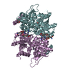
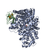
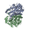
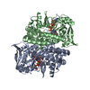
| Title | Crystal Structure of the Mouse-Muscle Adenylosuccinate Synthetase Ligated with GTP | ||||||
 Components Components | adenylosuccinate synthetase | ||||||
 Keywords Keywords | LIGASE / Purine biosynthesis / GTP-binding | ||||||
| Function / homology |  Function and homology information Function and homology informationPurine ribonucleoside monophosphate biosynthesis / AMP biosynthetic process / adenylosuccinate synthase / adenylosuccinate synthase activity / purine nucleotide metabolic process / aspartate metabolic process / cellular response to electrical stimulus / IMP metabolic process / 'de novo' AMP biosynthetic process / AMP salvage ...Purine ribonucleoside monophosphate biosynthesis / AMP biosynthetic process / adenylosuccinate synthase / adenylosuccinate synthase activity / purine nucleotide metabolic process / aspartate metabolic process / cellular response to electrical stimulus / IMP metabolic process / 'de novo' AMP biosynthetic process / AMP salvage / L-glutamine metabolic process / response to starvation / response to muscle activity / cellular response to xenobiotic stimulus / actin filament binding / GTPase activity / GTP binding / magnesium ion binding / membrane / identical protein binding / cytoplasm Similarity search - Function | ||||||
| Biological species |  | ||||||
| Method |  X-RAY DIFFRACTION / MOLECULAR REPLACEMENT / Resolution: 2.2 Å X-RAY DIFFRACTION / MOLECULAR REPLACEMENT / Resolution: 2.2 Å | ||||||
 Authors Authors | Iancu, C.V. / Borza, T. / Fromm, H.J. / Honzatko, R.B. | ||||||
 Citation Citation | Journal: J.Biol.Chem. / Year: 2002 Title: IMP, GTP, and 6-phosphoryl-IMP complexes of recombinant mouse muscle adenylosuccinate synthetase. Authors: Iancu, C.V. / Borza, T. / Fromm, H.J. / Honzatko, R.B. | ||||||
| History |
|
- Structure visualization
| Structure viewer | Molecule: MolmilJmol/JSmol |
|---|
- Downloads & links
-Download
| PDBx/mmCIF format | 1loo.cif.gz | 98.1 KB | Display | PDBx/mmCIF format |
|---|---|---|---|---|
| PDB format | pdb1loo.ent.gz | 74.4 KB | Display | PDB format |
| PDBx/mmJSON format | 1loo.json.gz | Tree view | PDBx/mmJSON format | |
| Others |  Other downloads Other downloads |
-Validation report
| Arichive directory | https://data.pdbj.org/pub/pdb/validation_reports/lo/1looftp://data.pdbj.org/pub/pdb/validation_reports/lo/1loo | HTTPS FTP |
|---|
-Related structure data
| Related structure data |  1iweC  1lnyC  1lonC  1j4bS S: Starting model for refinement C: citing same article ( |
|---|---|
| Similar structure data |


-Links
 PDBj
PDBj- Assembly
| Deposited unit | 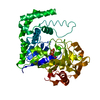
| ||||||||
|---|---|---|---|---|---|---|---|---|---|
| 1 | 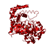
| ||||||||
| Unit cell |
| ||||||||
| Details | The biological assembly is a dimer. The asymmetric unit contains one monomer. The other monomer is generated by the symmetry operation: -y, -x, 1/2-z. |
-Components
| #1: Protein | Mass: 50321.301 Da / Num. of mol.: 1 Source method: isolated from a genetically manipulated source Source: (gene. exp.)  |
|---|---|
| #2: Chemical | ChemComp-GTP /   Mass: 523.180 Da / Num. of mol.: 1 / Source method: obtained synthetically / Formula: C10H16N5O14P3 / Comment: GTP, energy-carrying molecule*YM Mass: 523.180 Da / Num. of mol.: 1 / Source method: obtained synthetically / Formula: C10H16N5O14P3 / Comment: GTP, energy-carrying molecule*YM |
| #3: Water | ChemComp-HOH /  Mass: 18.015 Da / Num. of mol.: 100 / Source method: isolated from a natural source / Formula: H2O Mass: 18.015 Da / Num. of mol.: 100 / Source method: isolated from a natural source / Formula: H2O |
-Experimental details
-Experiment
| Experiment | Method: X-RAY DIFFRACTION / Number of used crystals: 1 |
|---|
- Sample preparation
| Crystal | Density Matthews: 2.41 Å3/Da / Density % sol: 48.87 % | ||||||||||||||||||||||||||||||||||||||||||||||||||||||||||||||||||||||
|---|---|---|---|---|---|---|---|---|---|---|---|---|---|---|---|---|---|---|---|---|---|---|---|---|---|---|---|---|---|---|---|---|---|---|---|---|---|---|---|---|---|---|---|---|---|---|---|---|---|---|---|---|---|---|---|---|---|---|---|---|---|---|---|---|---|---|---|---|---|---|---|
| Crystal grow | Temperature: 296 K / Method: vapor diffusion, hanging drop / pH: 7 Details: PEG 8000, magnesium acetate, pH 7, VAPOR DIFFUSION, HANGING DROP, temperature 296K | ||||||||||||||||||||||||||||||||||||||||||||||||||||||||||||||||||||||
| Crystal grow | *PLUS pH: 7.5 | ||||||||||||||||||||||||||||||||||||||||||||||||||||||||||||||||||||||
| Components of the solutions | *PLUS
|
-Data collection
| Diffraction | Mean temperature: 100 K |
|---|---|
| Diffraction source | Source: ROTATING ANODE / Type: RIGAKU / Wavelength: 1.5418 Å |
| Detector | Type: RIGAKU RAXIS IV / Detector: IMAGE PLATE / Date: Mar 19, 2002 / Details: osmic mirrors |
| Radiation | Protocol: SINGLE WAVELENGTH / Monochromatic (M) / Laue (L): M / Scattering type: x-ray |
| Radiation wavelength | Wavelength: 1.5418 Å / Relative weight: 1 |
| Reflection | Resolution: 2.2→65.9 Å / Num. all: 25715 / Num. obs: 21799 / % possible obs: 91.5 % / Observed criterion σ(F): 2 / Observed criterion σ(I): 2 / Redundancy: 4.9 % / Biso Wilson estimate: 47.4 Å2 / Rmerge(I) obs: 0.04 / Net I/σ(I): 20 |
| Reflection shell | Resolution: 2.2→2.28 Å / Redundancy: 4 % / Rmerge(I) obs: 0.255 / Mean I/σ(I) obs: 2.3 / Num. unique all: 2124 / % possible all: 89.5 |
| Reflection | *PLUS Num. obs: 25715 / % possible obs: 89.5 % / Num. measured all: 126081 / Rmerge(I) obs: 0.081 |
| Reflection shell | *PLUS Lowest resolution: 2.3 Å / % possible obs: 98.5 % / Rmerge(I) obs: 0.312 |
- Processing
| Software |
| |||||||||||||||||||||
|---|---|---|---|---|---|---|---|---|---|---|---|---|---|---|---|---|---|---|---|---|---|---|
| Refinement | Method to determine structure: MOLECULAR REPLACEMENT Starting model: 1J4B Resolution: 2.2→10 Å / Cross valid method: THROUGHOUT / σ(F): 0 / Stereochemistry target values: Engh & Huber
| |||||||||||||||||||||
| Displacement parameters | Biso mean: 46.9 Å2
| |||||||||||||||||||||
| Refine analyze |
| |||||||||||||||||||||
| Refinement step | Cycle: LAST / Resolution: 2.2→10 Å
| |||||||||||||||||||||
| Refine LS restraints |
| |||||||||||||||||||||
| LS refinement shell | Resolution: 2.2→2.34 Å / Rfactor Rfree error: 0.02
| |||||||||||||||||||||
| Refinement | *PLUS Lowest resolution: 10 Å / Num. reflection obs: 25407 / % reflection Rfree: 10 % / Rfactor Rfree: 0.276 / Rfactor Rwork: 0.244 | |||||||||||||||||||||
| Solvent computation | *PLUS | |||||||||||||||||||||
| Displacement parameters | *PLUS | |||||||||||||||||||||
| Refine LS restraints | *PLUS
| |||||||||||||||||||||
| LS refinement shell | *PLUS Rfactor Rfree: 0.382 / Rfactor Rwork: 0.338 |
