- PDB-4m9d: The Crystal structure of an adenylosuccinate synthetase from Baci... -
+
Open data
ID or keywords:
Loading...
-
Basic information
Entry
Database: PDB / ID: 4m9d
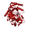
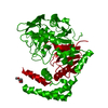
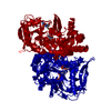
Title
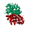
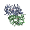
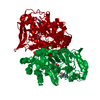
The Crystal structure of an adenylosuccinate synthetase from Bacillus anthracis str. Ames Ancestor in complex with AMP.
Components
Adenylosuccinate synthetase
Keywords
LIGASE / structural genomics / The Center for Structural Genomics of Infectious Diseases / CSGID / NIAID / National Institute of Allergy and Infectious Diseases
Function / homology
Function and homology information
adenylosuccinate synthase / adenylosuccinate synthase activity / IMP metabolic process / 'de novo' AMP biosynthetic process / GTP binding / magnesium ion binding / cytoplasm Similarity search - Function
Adenylosuccinate Synthetase; Chain A, domain 1 / Adenylosuccinate Synthetase, subunit A, domain 1 / Adenylosuccinate Synthetase; Chain A, domain 3 / Adenylosuccinate Synthetase, subunit A, domain 3 / Adenylosuccinate Synthetase, subunit A; domain 2 / Adenylosuccinate Synthetase, subunit A, domain 2 / Adenylosuccinate synthase, active site / Adenylosuccinate synthetase active site. / Adenylosuccinate synthase, GTP-binding site / Adenylosuccinate synthetase, domain 2 ...Adenylosuccinate Synthetase; Chain A, domain 1 / Adenylosuccinate Synthetase, subunit A, domain 1 / Adenylosuccinate Synthetase; Chain A, domain 3 / Adenylosuccinate Synthetase, subunit A, domain 3 / Adenylosuccinate Synthetase, subunit A; domain 2 / Adenylosuccinate Synthetase, subunit A, domain 2 / Adenylosuccinate synthase, active site / Adenylosuccinate synthetase active site. / Adenylosuccinate synthase, GTP-binding site / Adenylosuccinate synthetase, domain 2 / Adenylosuccinate synthetase, domain 3 / Adenylosuccinate synthetase GTP-binding site. / Adenylosuccinate synthetase / Adenylosuccinate synthetase, domain 1 / Adenylosuccinate synthetase / Adenylosuccinate synthetase / Alpha-Beta Complex / P-loop containing nucleoside triphosphate hydrolase / Orthogonal Bundle / 3-Layer(aba) Sandwich / Mainly Alpha / Alpha Beta Similarity search - Domain/homology
ADENOSINE MONOPHOSPHATE / FORMIC ACID / MALONATE ION / PHOSPHATE ION / Adenylosuccinate synthetase Similarity search - Component
In the structure databanks used in Yorodumi, some data are registered as the other names, "COVID-19 virus" and "2019-nCoV". Here are the details of the virus and the list of structure data.
Jan 31, 2019. EMDB accession codes are about to change! (news from PDBe EMDB page)
EMDB accession codes are about to change! (news from PDBe EMDB page)
The allocation of 4 digits for EMDB accession codes will soon come to an end. Whilst these codes will remain in use, new EMDB accession codes will include an additional digit and will expand incrementally as the available range of codes is exhausted. The current 4-digit format prefixed with “EMD-” (i.e. EMD-XXXX) will advance to a 5-digit format (i.e. EMD-XXXXX), and so on. It is currently estimated that the 4-digit codes will be depleted around Spring 2019, at which point the 5-digit format will come into force.
The EM Navigator/Yorodumi systems omit the EMD- prefix.
Related info.:Q: What is EMD? / ID/Accession-code notation in Yorodumi/EM Navigator
Yorodumi is a browser for structure data from EMDB, PDB, SASBDB, etc.
This page is also the successor to EM Navigator detail page, and also detail information page/front-end page for Omokage search.
The word "yorodu" (or yorozu) is an old Japanese word meaning "ten thousand". "mi" (miru) is to see.
Related info.:EMDB / PDB / SASBDB / Comparison of 3 databanks / Yorodumi Search / Aug 31, 2016. New EM Navigator & Yorodumi / Yorodumi Papers / Jmol/JSmol / Function and homology information / Changes in new EM Navigator and Yorodumi
 Movie
Movie Controller

 Yorodumi
 Open data
 Basic information
 Components
 Keywords
 Function and homology information
 X-RAY DIFFRACTION /
  Authors
 Citation
 Structure visualization
 Downloads & links
 Other downloads

 PDBj
PDBj


 Assembly


 Mass: 347.221 Da / Num. of mol.: 2 / Source method: obtained synthetically / Formula: C10H14N5O7P / Comment: AMP*YM
 Mass: 102.046 Da / Num. of mol.: 3 / Source method: obtained synthetically / Formula: C3H2O4
 Mass: 62.068 Da / Num. of mol.: 6 / Source method: obtained synthetically / Formula: C2H6O2
 Mass: 46.025 Da / Num. of mol.: 3 / Source method: obtained synthetically / Formula: CH2O2
 Mass: 94.971 Da / Num. of mol.: 1 / Source method: obtained synthetically / Formula: PO4
 Sample preparation
 / Beamline: 19-ID / Wavelength: 0.9786 Å
 Processing