 Movie
Movie Controller


+ Open data

- Basic information
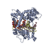
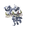
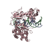
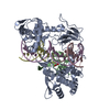
| Entry | Database: PDB / ID: 1a35 | ||||||
|---|---|---|---|---|---|---|---|
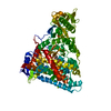
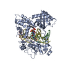
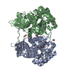
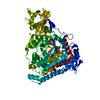
| Title | HUMAN TOPOISOMERASE I/DNA COMPLEX | ||||||
 Components Components |
| ||||||
 Keywords Keywords | ISOMERASE/DNA / TOPOISOMERASE I/DNA) / DNA / TOPOISOMERASE I / ISOMERASE-DNA COMPLEX | ||||||
| Function / homology |  Function and homology information Function and homology informationDNA topoisomerase / DNA topoisomerase type I (single strand cut, ATP-independent) activity / dense fibrillar component / cellular response to luteinizing hormone stimulus / programmed cell death / DNA binding, bending / supercoiled DNA binding / response to temperature stimulus / rRNA transcription / DNA topological change ...DNA topoisomerase / DNA topoisomerase type I (single strand cut, ATP-independent) activity / dense fibrillar component / cellular response to luteinizing hormone stimulus / programmed cell death / DNA binding, bending / supercoiled DNA binding / response to temperature stimulus / rRNA transcription / DNA topological change / animal organ regeneration / SUMOylation of DNA replication proteins / response to cAMP / response to gamma radiation / circadian regulation of gene expression / chromosome segregation / molecular condensate scaffold activity / circadian rhythm / P-body / chromatin DNA binding / protein-DNA complex / fibrillar center / chromosome / single-stranded DNA binding / double-stranded DNA binding / perikaryon / DNA replication / response to xenobiotic stimulus / RNA polymerase II cis-regulatory region sequence-specific DNA binding / chromatin remodeling / protein domain specific binding / protein serine/threonine kinase activity / chromatin binding / nucleolus / protein-containing complex binding / DNA binding / RNA binding / nucleoplasm / ATP binding / nucleus Similarity search - Function | ||||||
| Biological species |  Homo sapiens (human) Homo sapiens (human) | ||||||
| Method |  X-RAY DIFFRACTION / SYNCHROTRON / RIGID BODY REFINEMENT / Resolution: 2.5 Å X-RAY DIFFRACTION / SYNCHROTRON / RIGID BODY REFINEMENT / Resolution: 2.5 Å | ||||||
 Authors Authors | Redinbo, M.R. / Stewart, L. / Kuhn, P. / Champoux, J.J. / Hol, W.G. | ||||||
 Citation Citation | Journal: Science / Year: 1998 Title: Crystal structures of human topoisomerase I in covalent and noncovalent complexes with DNA. Authors: Redinbo, M.R. / Stewart, L. / Kuhn, P. / Champoux, J.J. / Hol, W.G. | ||||||
| History |
|
- Structure visualization
| Structure viewer | Molecule: MolmilJmol/JSmol |
|---|
- Downloads & links
-Download
| PDBx/mmCIF format | 1a35.cif.gz | 144.2 KB | Display | PDBx/mmCIF format |
|---|---|---|---|---|
| PDB format | pdb1a35.ent.gz | 105.6 KB | Display | PDB format |
| PDBx/mmJSON format | 1a35.json.gz | Tree view | PDBx/mmJSON format | |
| Others |  Other downloads Other downloads |
-Validation report
| Arichive directory | https://data.pdbj.org/pub/pdb/validation_reports/a3/1a35ftp://data.pdbj.org/pub/pdb/validation_reports/a3/1a35 | HTTPS FTP |
|---|
-Related structure data


-Links
 PDBj
PDBj


- Assembly
| Deposited unit | 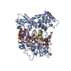
| ||||||||
|---|---|---|---|---|---|---|---|---|---|
| 1 |
| ||||||||
| Unit cell |
|
-Components
| #1: DNA chain | Mass: 6920.208 Da / Num. of mol.: 1 / Source method: obtained synthetically |
|---|---|
| #2: DNA chain | Mass: 7094.593 Da / Num. of mol.: 1 / Source method: obtained synthetically |
| #3: Protein | Mass: 70022.859 Da / Num. of mol.: 1 / Fragment: CORE DOMAIN AND C-TERMINAL DOMAIN / Mutation: Y723F Source method: isolated from a genetically manipulated source Source: (gene. exp.) Homo sapiens (human) / Cellular location: NUCLEUS / Production host:   Spodoptera frugiperda (fall armyworm) / References: UniProt: P11387, EC: 5.99.1.2 Spodoptera frugiperda (fall armyworm) / References: UniProt: P11387, EC: 5.99.1.2 |
| #4: Water | ChemComp-HOH /  Mass: 18.015 Da / Num. of mol.: 273 / Source method: isolated from a natural source / Formula: H2O Mass: 18.015 Da / Num. of mol.: 273 / Source method: isolated from a natural source / Formula: H2O |
| Compound details | RESIDUES 215-635 OF CHAIN A COMPRISE THE CORE DOMAIN OF THE MOLECULE. RESIDUES 715-765 OF CHAIN A ...RESIDUES 215-635 OF CHAIN A COMPRISE THE CORE DOMAIN OF THE MOLECULE. RESIDUES 715-765 OF CHAIN A COMPRISE THE C-TERMINAL DOMAIN. 1-22 OF CHAIN C COMPRISE THE SCISSILE STRAND OF THE DNA, 101-122 OF CHAIN D THE INTACT DNA STRAND. NOTE THAT THE LINKER DOMAIN (636-714) IS NOT PRESENT IN THE CONSTRUCT OF THIS RECONSTITU |
-Experimental details
-Experiment
| Experiment | Method: X-RAY DIFFRACTION / Number of used crystals: 1 |
|---|
- Sample preparation
| Crystal | Density Matthews: 2.01 Å3/Da / Density % sol: 38.91 % | |||||||||||||||||||||||||||||||||||||||||||||||||||||||||||||||||||||||||||||||||||||||||||
|---|---|---|---|---|---|---|---|---|---|---|---|---|---|---|---|---|---|---|---|---|---|---|---|---|---|---|---|---|---|---|---|---|---|---|---|---|---|---|---|---|---|---|---|---|---|---|---|---|---|---|---|---|---|---|---|---|---|---|---|---|---|---|---|---|---|---|---|---|---|---|---|---|---|---|---|---|---|---|---|---|---|---|---|---|---|---|---|---|---|---|---|---|
| Crystal grow | Temperature: 295 K / Method: vapor diffusion, sitting drop / pH: 7.7 Details: pH 7.70, VAPOR DIFFUSION, SITTING DROP, temperature 295.00K | |||||||||||||||||||||||||||||||||||||||||||||||||||||||||||||||||||||||||||||||||||||||||||
| Components of the solutions |
| |||||||||||||||||||||||||||||||||||||||||||||||||||||||||||||||||||||||||||||||||||||||||||
| Crystal grow | *PLUS Temperature: 22 ℃ / pH: 7.5 | |||||||||||||||||||||||||||||||||||||||||||||||||||||||||||||||||||||||||||||||||||||||||||
| Components of the solutions | *PLUS
|
-Data collection
| Diffraction | Mean temperature: 100 K |
|---|---|
| Diffraction source | Source: SYNCHROTRON / Site: NSLS  / Beamline: X4A / Beamline: X4A |
| Detector | Type: FUJI / Detector: IMAGE PLATE / Date: Jun 1, 1996 |
| Radiation | Protocol: SINGLE WAVELENGTH / Monochromatic (M) / Laue (L): M / Scattering type: x-ray |
| Radiation wavelength | Relative weight: 1 |
| Reflection | Resolution: 2.5→20 Å / Num. obs: 18834 / % possible obs: 83.9 % / Observed criterion σ(I): 2 / Redundancy: 4.2 % / Rmerge(I) obs: 0.077 |
| Reflection shell | Resolution: 2.5→2.6 Å / Redundancy: 4.2 % / Rmerge(I) obs: 0.269 / % possible all: 87.7 |
| Reflection | *PLUS Highest resolution: 2.5 Å / Lowest resolution: 20 Å / % possible obs: 83.9 % / Observed criterion σ(I): 2 / Redundancy: 4.2 % / Num. measured all: 78779 |
| Reflection shell | *PLUS Highest resolution: 2.5 Å / Lowest resolution: 2.6 Å / % possible obs: 87.7 % / Redundancy: 4.2 % |
- Processing
| Software |
| ||||||||||||||||||||||||||||||||||||||||||||||||||||||||||||
|---|---|---|---|---|---|---|---|---|---|---|---|---|---|---|---|---|---|---|---|---|---|---|---|---|---|---|---|---|---|---|---|---|---|---|---|---|---|---|---|---|---|---|---|---|---|---|---|---|---|---|---|---|---|---|---|---|---|---|---|---|---|
| Refinement | Method to determine structure: RIGID BODY REFINEMENT Starting model: RECONSTITUTED HUMAN TOPOISOMERASE I COVALENT COMPLEX WITH 22 BASE PAIR DUPLEX DNA Resolution: 2.5→20 Å / Data cutoff high absF: 10000000 / Data cutoff low absF: 0.001 / Cross valid method: THROUGHOUT / σ(F): 2
| ||||||||||||||||||||||||||||||||||||||||||||||||||||||||||||
| Displacement parameters |
| ||||||||||||||||||||||||||||||||||||||||||||||||||||||||||||
| Refinement step | Cycle: LAST / Resolution: 2.5→20 Å
| ||||||||||||||||||||||||||||||||||||||||||||||||||||||||||||
| Refine LS restraints |
| ||||||||||||||||||||||||||||||||||||||||||||||||||||||||||||
| LS refinement shell | Resolution: 2.5→2.59 Å / Total num. of bins used: 10
| ||||||||||||||||||||||||||||||||||||||||||||||||||||||||||||
| Xplor file |
| ||||||||||||||||||||||||||||||||||||||||||||||||||||||||||||
| Software | *PLUS Name: X-PLOR / Version: 3.1 / Classification: refinement | ||||||||||||||||||||||||||||||||||||||||||||||||||||||||||||
| Refinement | *PLUS Highest resolution: 2.5 Å / Lowest resolution: 20 Å / σ(F): 2 / % reflection Rfree: 7 % / Rfactor obs: 0.209 | ||||||||||||||||||||||||||||||||||||||||||||||||||||||||||||
| Solvent computation | *PLUS | ||||||||||||||||||||||||||||||||||||||||||||||||||||||||||||
| Displacement parameters | *PLUS | ||||||||||||||||||||||||||||||||||||||||||||||||||||||||||||
| Refine LS restraints | *PLUS
| ||||||||||||||||||||||||||||||||||||||||||||||||||||||||||||
| LS refinement shell | *PLUS % reflection Rfree: 6 % |
