[English] 日本語
Yorodumi
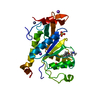
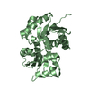
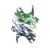
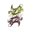
- PDB-1dqt: THE CRYSTAL STRUCTURE OF MURINE CTLA4 (CD152) -

+
Open data


ID or keywords:

Loading...

-
Basic information

Entry
Database: PDB / ID: 1dqt
TitleTHE CRYSTAL STRUCTURE OF MURINE CTLA4 (CD152)
ComponentsCYTOTOXIC T LYMPHOCYTE ASSOCIATED ANTIGEN 4
KeywordsIMMUNE SYSTEM / immunoglobulin variable domain-like beta-sandwich / homodimer
Function / homology
Function and homology information


CTLA4 inhibitory signaling / protein complex involved in cell adhesion / negative regulation of regulatory T cell differentiation / clathrin-coated endocytic vesicle / negative regulation of B cell proliferation / negative regulation of T cell proliferation / B cell receptor signaling pathway / T cell receptor signaling pathway / adaptive immune response / positive regulation of apoptotic process ...CTLA4 inhibitory signaling / protein complex involved in cell adhesion / negative regulation of regulatory T cell differentiation / clathrin-coated endocytic vesicle / negative regulation of B cell proliferation / negative regulation of T cell proliferation / B cell receptor signaling pathway / T cell receptor signaling pathway / adaptive immune response / positive regulation of apoptotic process / external side of plasma membrane / DNA damage response / perinuclear region of cytoplasm / Golgi apparatus / plasma membrane
Similarity search - Function
Cytotoxic T-lymphocyte antigen 4 / Cytotoxic T-lymphocyte protein 4/CD28 / Immunoglobulin V-Type / Immunoglobulin V-set domain / Immunoglobulin V-set domain / Immunoglobulin subtype / Immunoglobulin / Ig-like domain profile. / Immunoglobulin-like domain / Immunoglobulin-like domain superfamily ...Cytotoxic T-lymphocyte antigen 4 / Cytotoxic T-lymphocyte protein 4/CD28 / Immunoglobulin V-Type / Immunoglobulin V-set domain / Immunoglobulin V-set domain / Immunoglobulin subtype / Immunoglobulin / Ig-like domain profile. / Immunoglobulin-like domain / Immunoglobulin-like domain superfamily / Immunoglobulins / Immunoglobulin-like fold / Immunoglobulin-like / Sandwich / Mainly Beta
Similarity search - Domain/homology
Cytotoxic T-lymphocyte protein 4
Similarity search - Component
Biological speciesMus musculus (house mouse)
MethodX-RAY DIFFRACTION / SYNCHROTRON / Resolution: 2 Å
AuthorsOstrov, D.A. / Shi, W. / Schwartz, J.C. / Almo, S.C. / Nathenson, S.G.
CitationJournal: Science / Year: 2000
Title: Structure of murine CTLA-4 and its role in modulating T cell responsiveness.
Authors: Ostrov, D.A. / Shi, W. / Schwartz, J.C. / Almo, S.C. / Nathenson, S.G.
History
DepositionJan 5, 2000Deposition site: RCSB / Processing site: RCSB
Revision 1.0Oct 27, 2000Provider: repository / Type: Initial release
Revision 1.1Apr 27, 2008Group: Version format compliance
Revision 1.2Jul 13, 2011Group: Version format compliance
Revision 1.3Jan 31, 2018Group: Experimental preparation / Category: exptl_crystal_grow / Item: _exptl_crystal_grow.temp

-
Structure visualization

Structure viewerMolecule:
MolmilJmol/JSmol

Downloads & links

-
Assembly

Deposited unit
A: CYTOTOXIC T LYMPHOCYTE ASSOCIATED ANTIGEN 4
B: CYTOTOXIC T LYMPHOCYTE ASSOCIATED ANTIGEN 4
C: CYTOTOXIC T LYMPHOCYTE ASSOCIATED ANTIGEN 4
D: CYTOTOXIC T LYMPHOCYTE ASSOCIATED ANTIGEN 4
hetero molecules


Theoretical massNumber of molelcules
Total (without water)51,9149
Polymers51,7104
Non-polymers2045
Water7,782432
1
A: CYTOTOXIC T LYMPHOCYTE ASSOCIATED ANTIGEN 4
B: CYTOTOXIC T LYMPHOCYTE ASSOCIATED ANTIGEN 4
hetero molecules


Theoretical massNumber of molelcules
Total (without water)25,9885
Polymers25,8552
Non-polymers1333
Water362
TypeNameSymmetry operationNumber
identity operation1_555x,y,z1
Buried area1970 Å2
ΔGint-27 kcal/mol
Surface area11600 Å2
MethodPISA
2
C: CYTOTOXIC T LYMPHOCYTE ASSOCIATED ANTIGEN 4
D: CYTOTOXIC T LYMPHOCYTE ASSOCIATED ANTIGEN 4
hetero molecules


Theoretical massNumber of molelcules
Total (without water)25,9264
Polymers25,8552
Non-polymers712
Water362
TypeNameSymmetry operationNumber
identity operation1_555x,y,z1
Buried area1820 Å2
ΔGint-29 kcal/mol
Surface area11480 Å2
MethodPISA
3


  • Idetical with deposited unit
  • defined by software
TypeNameSymmetry operationNumber
identity operation1_555x,y,z1
Buried area5610 Å2
ΔGint-78 kcal/mol
Surface area21270 Å2
MethodPISA
Unit cell
Length a, b, c (Å)112.85, 112.85, 102.504
Angle α, β, γ (deg.)90.0, 90.0, 90.0
Int Tables number96
Space group name H-MP43212

-
Components

#1: Protein
CYTOTOXIC T LYMPHOCYTE ASSOCIATED ANTIGEN 4 / CTLA4


Mass: 12927.545 Da / Num. of mol.: 4 / Fragment: EXTRACELLULAR FRAGMENT
Source method: isolated from a genetically manipulated source
Source: (gene. exp.) Mus musculus (house mouse) / Plasmid: PET3 / Production host: Escherichia coli (E. coli) / References: UniProt: P09793
#2: Chemical
ChemComp-CL / CHLORIDE ION / Chloride


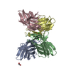
Mass: 35.453 Da / Num. of mol.: 4 / Source method: obtained synthetically / Formula: Cl
#3: Chemical ChemComp-EDO / 1,2-ETHANEDIOL / ETHYLENE GLYCOL / Ethylene glycol


Mass: 62.068 Da / Num. of mol.: 1 / Source method: obtained synthetically / Formula: C2H6O2
#4: Water ChemComp-HOH / water / Water


Mass: 18.015 Da / Num. of mol.: 432 / Source method: isolated from a natural source / Formula: H2O

-
Experimental details

-
Experiment

ExperimentMethod: X-RAY DIFFRACTION / Number of used crystals: 1

-
Sample preparation

CrystalDensity Matthews: 3.15 Å3/Da / Density % sol: 61 %
Crystal growTemperature: 291 K / Method: vapor diffusion, hanging drop / pH: 6.5
Details: 18% PEG 6000, 2.0 M NaCl, pH 6.5, VAPOR DIFFUSION, HANGING DROP, temperature 18.0K
Crystal
*PLUS
Density % sol: 58 %
Crystal grow
*PLUS
Temperature: 18 ℃
Components of the solutions
*PLUS
IDConc.Common nameCrystal-IDSol-IDChemical formula
110 mg/mlprotein1drop
220 %PEG60001reservoir
32.0 M1reservoirNaCl

-
Data collection

DiffractionMean temperature: 100 K
Diffraction sourceSource: SYNCHROTRON / Site: NSLS / Beamline: X9B / Wavelength: 0.9795
DetectorType: MARRESEARCH / Detector: AREA DETECTOR / Date: Jun 28, 1999
RadiationProtocol: SINGLE WAVELENGTH / Monochromatic (M) / Laue (L): M / Scattering type: x-ray
Radiation wavelengthWavelength: 0.9795 Å / Relative weight: 1
ReflectionResolution: 2→10 Å / Num. all: 44587 / Num. obs: 44587 / % possible obs: 100 % / Observed criterion σ(F): 0 / Observed criterion σ(I): 0 / Redundancy: 13.7 % / Biso Wilson estimate: 16.4 Å2 / Rsym value: 0.046 / Net I/σ(I): 36.5
Reflection shellResolution: 2→2.07 Å / Redundancy: 5.8 % / Num. unique all: 4206 / Rsym value: 0.057 / % possible all: 95.7
Reflection
*PLUS
% possible obs: 99.4 % / Num. measured all: 301486 / Rmerge(I) obs: 0.062
Reflection shell
*PLUS
% possible obs: 95.7 % / Rmerge(I) obs: 0.363 / Mean I/σ(I) obs: 5.7

-
Processing

Software
NameClassification
DENZOdata reduction
SCALEPACKdata scaling
SOLVEphasing
CNSrefinement
RefinementResolution: 2→10 Å / Rfactor Rfree error: 0.004 / Data cutoff high rms absF: 309707 / Isotropic thermal model: restrained / Cross valid method: THROUGHOUT / σ(F): 2 / σ(I): 1.4 / Stereochemistry target values: Engh & Huber / Details: Used weighted full matrix least squares procedure.
RfactorNum. reflection% reflectionSelection details
Rfree0.2543 3965 10 %RANDOM
Rwork0.2106 ---
all0.228 41889 --
obs0.2217 39329 93.4 %-
Displacement parametersBiso mean: 31.3 Å2
Baniso -1Baniso -2Baniso -3
1-5.21 Å2--
2--5.21 Å2-
3---10.42 Å2
Refine analyze
FreeObs
Luzzati coordinate error0.3 Å0.24 Å
Luzzati d res low-5 Å
Luzzati sigma a0.25 Å0.22 Å
Refinement stepCycle: LAST / Resolution: 2→10 Å
ProteinNucleic acidLigandSolventTotal
Num. atoms3624 0 8 432 4064
Refine LS restraints
Refine-IDTypeDev ideal
X-RAY DIFFRACTIONc_bond_d0.009143
X-RAY DIFFRACTIONc_angle_deg1.52217
X-RAY DIFFRACTIONc_dihedral_angle_d27.7
X-RAY DIFFRACTIONc_improper_angle_d0.92
LS refinement shellResolution: 2→2.13 Å / Rfactor Rfree error: 0.012 / Total num. of bins used: 6
RfactorNum. reflection% reflection
Rfree0.309 686 10.5 %
Rwork0.292 5822 -
obs--87.9 %
Software
*PLUS
Name: CNS / Classification: refinement
Refinement
*PLUS
Highest resolution: 2 Å / Lowest resolution: 10 Å / σ(F): 2 / % reflection Rfree: 10 %
Solvent computation
*PLUS
Displacement parameters
*PLUS
Biso mean: 31.3 Å2
Refine LS restraints
*PLUS
Refine-IDTypeDev ideal
X-RAY DIFFRACTIONc_dihedral_angle_d
X-RAY DIFFRACTIONc_dihedral_angle_deg27.7
X-RAY DIFFRACTIONc_improper_angle_d
X-RAY DIFFRACTIONc_improper_angle_deg0.92
LS refinement shell
*PLUS
Rfactor Rfree: 0.309 / % reflection Rfree: 10.5 % / Rfactor Rwork: 0.292

+
About Yorodumi

-
News

-
Feb 9, 2022. New format data for meta-information of EMDB entries

New format data for meta-information of EMDB entries

  • Version 3 of the EMDB header file is now the official format.
  • The previous official version 1.9 will be removed from the archive.

Related info.:EMDB header

External links:wwPDB to switch to version 3 of the EMDB data model

-
Aug 12, 2020. Covid-19 info

Covid-19 info

URL: https://pdbj.org/emnavi/covid19.php

New page: Covid-19 featured information page in EM Navigator.

Related info.:Covid-19 info / Mar 5, 2020. Novel coronavirus structure data

+
Mar 5, 2020. Novel coronavirus structure data

Novel coronavirus structure data

Related info.:Yorodumi Speices / Aug 12, 2020. Covid-19 info

External links:COVID-19 featured content - PDBj / Molecule of the Month (242):Coronavirus Proteases

+
Jan 31, 2019. EMDB accession codes are about to change! (news from PDBe EMDB page)

EMDB accession codes are about to change! (news from PDBe EMDB page)

  • The allocation of 4 digits for EMDB accession codes will soon come to an end. Whilst these codes will remain in use, new EMDB accession codes will include an additional digit and will expand incrementally as the available range of codes is exhausted. The current 4-digit format prefixed with “EMD-” (i.e. EMD-XXXX) will advance to a 5-digit format (i.e. EMD-XXXXX), and so on. It is currently estimated that the 4-digit codes will be depleted around Spring 2019, at which point the 5-digit format will come into force.
  • The EM Navigator/Yorodumi systems omit the EMD- prefix.

Related info.:Q: What is EMD? / ID/Accession-code notation in Yorodumi/EM Navigator

External links:EMDB Accession Codes are Changing Soon! / Contact to PDBj

+
Jul 12, 2017. Major update of PDB

Major update of PDB

  • wwPDB released updated PDB data conforming to the new PDBx/mmCIF dictionary.
  • This is a major update changing the version number from 4 to 5, and with Remediation, in which all the entries are updated.
  • In this update, many items about electron microscopy experimental information are reorganized (e.g. em_software).
  • Now, EM Navigator and Yorodumi are based on the updated data.

External links:wwPDB Remediation / Enriched Model Files Conforming to OneDep Data Standards Now Available in the PDB FTP Archive

-
Yorodumi

Thousand views of thousand structures

  • Yorodumi is a browser for structure data from EMDB, PDB, SASBDB, etc.
  • This page is also the successor to EM Navigator detail page, and also detail information page/front-end page for Omokage search.
  • The word "yorodu" (or yorozu) is an old Japanese word meaning "ten thousand". "mi" (miru) is to see.

Related info.:EMDB / PDB / SASBDB / Comparison of 3 databanks / Yorodumi Search / Aug 31, 2016. New EM Navigator & Yorodumi / Yorodumi Papers / Jmol/JSmol / Function and homology information / Changes in new EM Navigator and Yorodumi

Read more